File:Zebrafish day 1 SEM.jpg
Original file (1,000 × 750 pixels, file size: 130 KB, MIME type: image/jpeg)
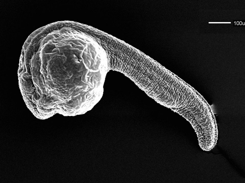
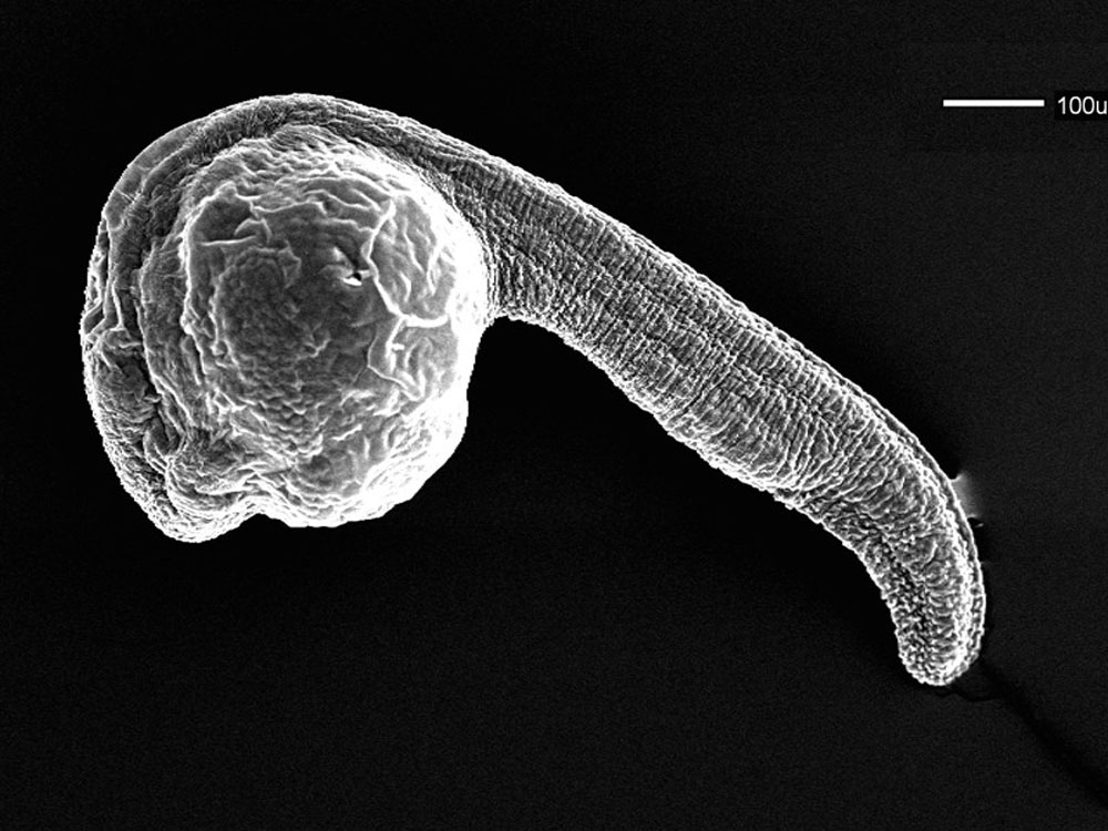
Zebrafish Day 1 SEM
Scanning EM of a 24 hr (prim-5) zebrafish embryo.
Zebrafish were chemically fixed critically point dried, and sputter coated with gold/palladium. This image is part of a series taken by Bryan Crawford while he was at the University of Washington. They are part of the Zebrafish--The Living Laboratory CD made available by Mark Cooper and described in Methods in Cell Biology Volume 77, 2004, Pages 439-457.
Original image name: 12661.jpg http://www.cellimagelibrary.org/images/12661
Licensing: Attribution Non-Commercial Share Alike:This image is licensed under a Creative Commons Attribution, Non-Commercial Share Alike License.
File history
Yi efo/eka'e gwa ebo wo le nyangagi wuncin ye kamina wunga tinya nan
| Gwalagizhi | Nyangagi | Dimensions | User | Comment | |
|---|---|---|---|---|---|
| current | 08:16, 26 April 2011 | | 1,000 × 750 (130 KB) | S8600021 (talk | contribs) | ==Zebrafish Day 1 SEM== Scanning EM of a 24 hr (prim-5) zebrafish embryo. Zebrafish were chemically fixed critically point dried, and sputter coated with gold/palladium. This image is part of a series taken by Bryan Crawford while he was at the Univer |
You cannot overwrite this file.
File usage
The following page uses this file: