File:Buell-plate01.jpg
Original file (1,221 × 1,500 pixels, file size: 236 KB, MIME type: image/jpeg)
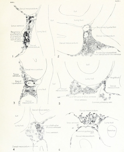
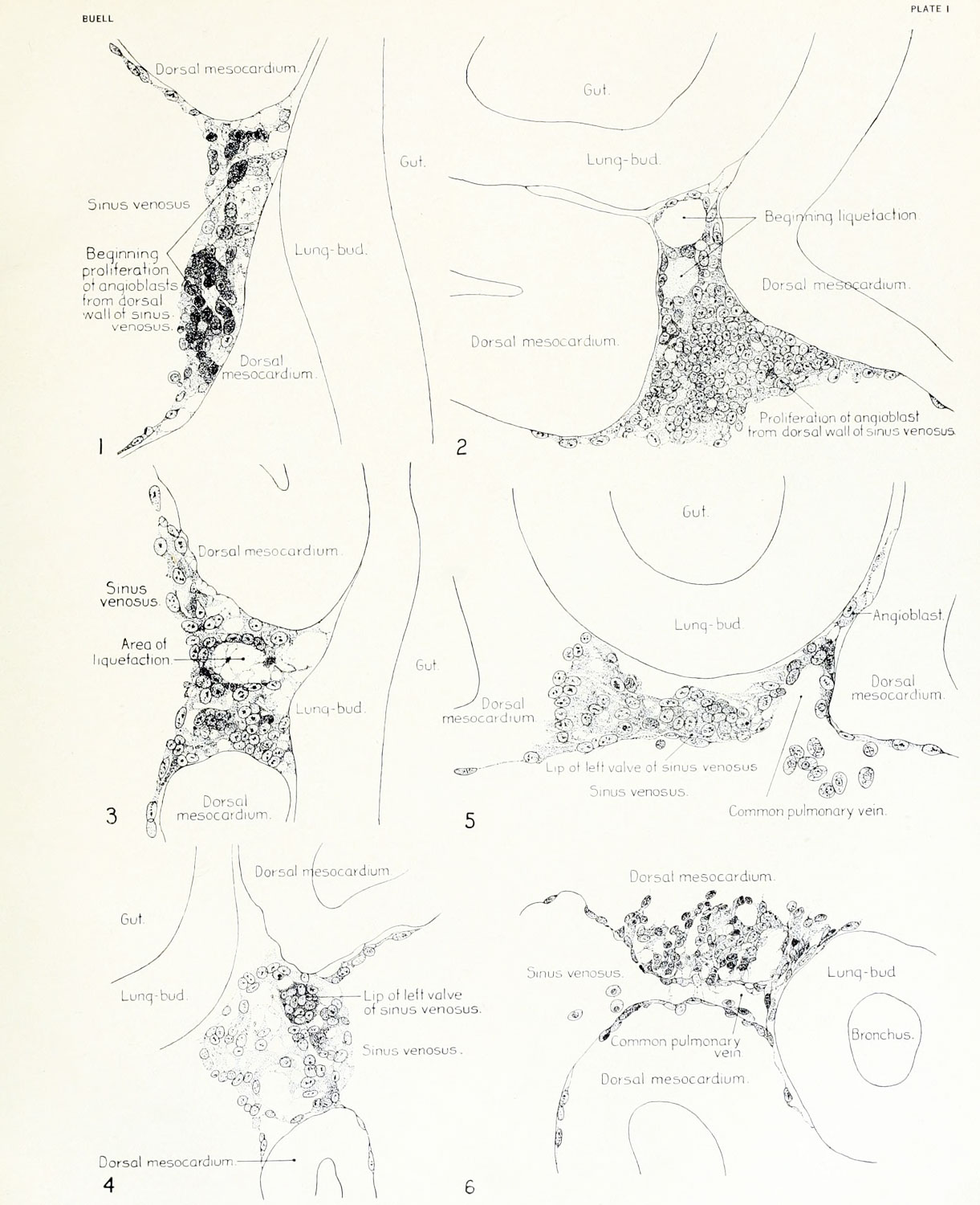
Fig. 1 - 20-somite chick
Median-sagittal section (10 micron in thickness) through tip of lung-bud of a 20-somite chick, 40 hours' incubation; hematoxylin and eosin slain, series B. Angioblasts, forerunners of pulmonary system, are seen proliferating from and near dorsal endothelial wall of sinus venosus. They extend dorsally toward the ventral surface of the gut, which shows a slight ventral swelling - the primary lung rudiment. This is about the earliest stage in which there is any evidence of the formation of a pulmonary vascular system.
Fig. 2 - 21-somite chick
Cross-section (10 micron) through tip of primary lung rudiment of a 21-somite chick, 48 hours' incubation; carmine stain, series 0. A slightly later stage in the angioblastic proliferation from the dorsal sinus wall.
The right portion of the cell-mass has begun to form a matted group of cells, (lie tip of the left valve of the sinus venosus. The left portion of the proliferation shows signs of liquefaction by which the common pulmonary vein is formed.
Fig. 3 - 23-somite chick
Sagittal section (10 micron) through left third of proliferation of angioblasts, to show process of liquefaction extending toward sinus venosus. It is about to open into the sinus, thus forming the common pulmonary vein.
Hematoxylin and eosin stain; chick of 23 somites, 48 hours' incubation, series D.
Fig. 4 - 22-somite chick
Median-sagittal section (10 micron) through right two-thirds of mass of angioblasts, to show matting together of cells to form left sinus valve tip.
The pulmonary vein is not established.
This embryo (22 somites, 44 hours' incubation) is slightly younger than that shown in figure 3.
Hematoxylin and eosin stain; series II.
Fig. 5 - 24-somite chick
Cross-section (10 micron) through tip of primary lung rudiment of a chick of 24 somites, 48 hours' incubation;
carmine stain, series E. The right two-thirds of the mass of angioblasts has formed the tip of the left valve of the sinus.
The common pulmonary vein is now established, opening into the sinus to the left of the valve. Angioblasts can be seen spreading over the ventral surface of the gut. This is the earliest stage in which the common vein is complete.
Fig. 6 - 31-somite chick
Sagittal section (15 micron) through plane of common pulmonary vein, showing it complete, from the sinus venosus to tip of lung rudiment.
There is no pulmonary circulation at this stage Angioblasts can be seen over the surface of the lung-bud.
Embryo of 31 somites, 50 hours' incubation; hematoxylin and eosin stain; series X.
File history
Yi efo/eka'e gwa ebo wo le nyangagi wuncin ye kamina wunga tinya nan
| Gwalagizhi | Nyangagi | Dimensions | User | Comment | |
|---|---|---|---|---|---|
| current | 16:38, 23 Ocak 2013 | | 1,221 × 1,500 (236 KB) | Z8600021 (talk | contribs) | JP2 scan |
| 11:54, 29 March 2011 |  | 1,028 × 1,332 (160 KB) | S8600021 (talk | contribs) | Cross-section (10 m) through tip of primary lung rudiment of a 21-somite chick, 48 hours' incubation; carmine stain, series 0. A slightly later stage in the angioblastic proliferation from the dorsal sinus wall. The right portion of the cell-mass has be |
You cannot overwrite this file.
File usage
The following page uses this file: