File:Lymph node histology 01.jpg
From Embryology
No higher resolution available.
Lymph_node_histology_01.jpg (600 × 400 pixels, file size: 61 KB, MIME type: image/jpeg)
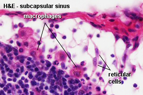
Lymph Node Histology
- "Reticular cells (and reticular fibres) form a delicate network between the capsule and trabeculae. Only their large and light nuclei are easily visible in the microscope. The cytoplasm of reticular cells is only weakly eosinophilic. Lymphocytes and macrophages are housed in the network of reticular cells and the reticular fibres formed by them. The processes of reticular cells and reticular fibres extend into and criss-cross within the sinuses."
Original file name: Lyn44he.jpg http://www.lab.anhb.uwa.edu.au/mb140/CorePages/Lymphoid1/lymph1.htm#Lymph
Lymph node histology 01.jpg
File history
Yi efo/eka'e gwa ebo wo le nyangagi wuncin ye kamina wunga tinya nan
| Gwalagizhi | Nyangagi | Dimensions | User | Comment | |
|---|---|---|---|---|---|
| current | 18:32, 25 February 2012 | | 600 × 400 (61 KB) | Z8600021 (talk | contribs) | increase image size |
| 08:57, 14 February 2011 | 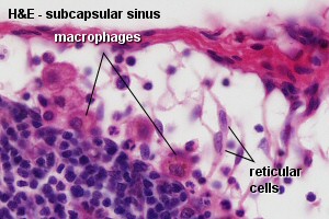 | 300 × 200 (25 KB) | S8600021 (talk | contribs) | ==Lymph Node Histology== :"Reticular cells (and reticular fibres) form a delicate network between the capsule and trabeculae. Only their large and light nuclei are easily visible in the microscope. The cytoplasm of reticular cells is only weakly eosinoph |
You cannot overwrite this file.