File:Neural- cortex Cajal drawing 01.jpg
From Embryology
Size of this preview: 518 × 599 pixels.
Original file (800 × 925 pixels, file size: 154 KB, MIME type: image/jpeg)
Cortex Historic Drawing by Cajal
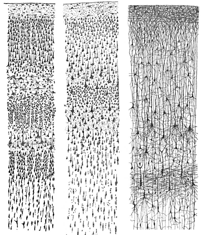
Cajal's historic drawing of histologically stained human cortex sections. Surface of the cortex at the top of image.
- Left - adult human visual cortex. (Nissl-stain)
- Middle - adult human motor cortex (Nissl-stain)
- Right: - infant human (1½ month) (Golgi-stain)
Histology Stains
- Nissl stain - shows the cell bodies of neurons
- Golgi stain - shows the dendrites and axons of a random subset of neurons. Developed by Camillo Golgi ((1843 – 1926)
Santiago Ramon y Cajal (1852 – 1934) was a Spanish histologist and neuroscientist. He also won the Nobel Prize in Physiology or Medicine (1906).
File history
Yi efo/eka'e gwa ebo wo le nyangagi wuncin ye kamina wunga tinya nan
| Gwalagizhi | Nyangagi | Dimensions | User | Comment | |
|---|---|---|---|---|---|
| current | 07:42, 15 December 2010 | | 800 × 925 (154 KB) | S8600021 (talk | contribs) | ==Cortex Historic Drawing by Cajal== Cajal's historic drawing of histologically stained human cortex sections. Surface of the cortex at the top of image. * Left - adult human visual cortex. (Nissl-stain) * Middle - adult human motor cortex (Nissl-stai |
You cannot overwrite this file.
File usage
The following 4 pages use this file:


