File:Gray0556.jpg
Gray0556.jpg (600 × 533 pixels, file size: 86 KB, MIME type: image/jpeg)
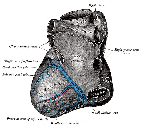
Coronary Circulation - Veins
Heart viewed from the base and diaphragmatic surface.
Most of the veins of the heart open into the coronary sinus. This is a wide venous channel about 2.25 cm. in length situated in the posterior part of the coronary sulcus, and covered by muscular fibers from the left atrium.
It ends in the right atrium between the opening of the inferior vena cava and the atrioventricular aperture, its orifice being guarded by a semilunar valve, the valve of the coronary sinus (valve of Thebesius).
Tributaries
Its tributaries are the great, small, and middle cardiac veins, the posterior vein of the left ventricle, and the oblique vein of the left atrium, all of which, except the last, are provided with valves at their orifices.
Cardiac veins not ending in the coronary sinus
- anterior cardiac veins - comprising three or four small vessels which collect blood from the front of the right ventricle and open into the right atrium; the right marginal vein frequently opens into the right atrium, and is therefore sometimes regarded as belonging to this group
- smallest cardiac veins (veins of Thebesius) - consisting of a number of minute veins which arise in the muscular wall of the heart; the majority open into the atria, but a few end in the ventricles.
(text modified from Gray's 1918 Anatomy)
File history
Yi efo/eka'e gwa ebo wo le nyangagi wuncin ye kamina wunga tinya nan
| Gwalagizhi | Nyangagi | Dimensions | User | Comment | |
|---|---|---|---|---|---|
| current | 11:18, 16 October 2010 |  | 600 × 533 (86 KB) | S8600021 (talk | contribs) |
You cannot overwrite this file.
File usage
The following 2 pages use this file:
