File:Cervical ectopic ultrasound.jpg
Cervical_ectopic_ultrasound.jpg (800 × 541 pixels, file size: 62 KB, MIME type: image/jpeg)
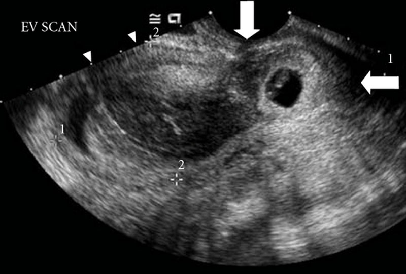
Cervical Ectopic Pregnancy
A gestational sac with a small embryonic pole with a fetal heartbeat of 122bpm located in the cervix below the scar of the previous cesarean section (vertical arrow). Cervix was closed, enlarged, and tender (horizontal arrow). Estimated gestational age based on last menstrual period was 6 weeks and 6 days.
Reference
Mohebbi MR, Rosenkrans KA, Luebbert EE, Hunt TT & Jung MJ. (2011). Ectopic pregnancy in the cervix: a case report. Case Rep Med , 2011, 858241. PMID: 22110520 DOI.
Copyright
This is an open access article distributed under the Creative Commons Attribution License, which permits unrestricted use, distribution, and reproduction in any medium, provided the original work is properly cited.
Original file name Figure 1 CRIM2011-858241.001.jpg (image cropped and resized)
Cite this page: Hill, M.A. (2024, June 16) Embryology Cervical ectopic ultrasound.jpg. Retrieved from https://embryology.med.unsw.edu.au/embryology/index.php/File:Cervical_ectopic_ultrasound.jpg
- © Dr Mark Hill 2024, UNSW Embryology ISBN: 978 0 7334 2609 4 - UNSW CRICOS Provider Code No. 00098G
File history
Click on a date/time to view the file as it appeared at that time.
| Date/Time | Thumbnail | Dimensions | User | Comment | |
|---|---|---|---|---|---|
| current | 13:55, 18 January 2012 | | 800 × 541 (62 KB) | S8600021 (talk | contribs) | CRIM2011-858241.001.jpg |
You cannot overwrite this file.
File usage
The following 2 pages use this file: