File:Mouse-sciatic nerve Schwann cell.jpg
Original file (957 × 1,050 pixels, file size: 339 KB, MIME type: image/jpeg)
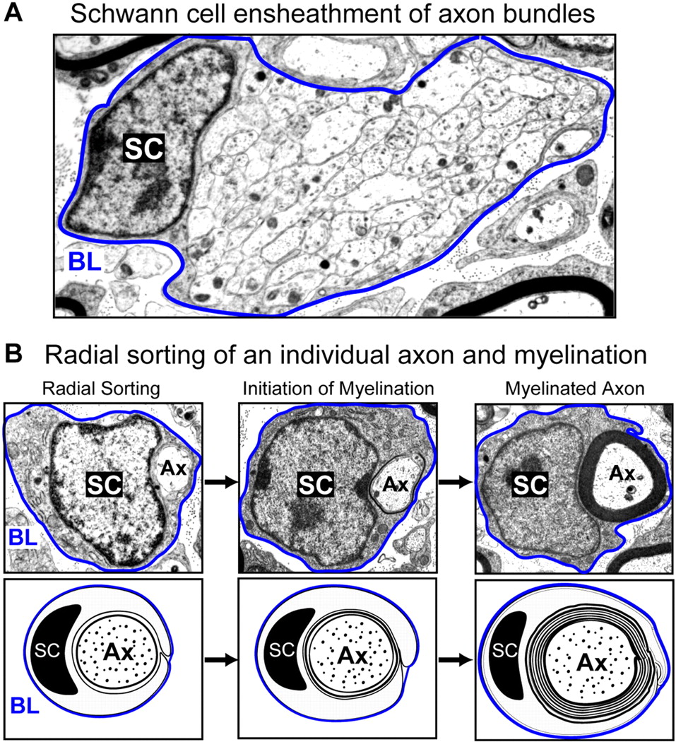
Illustration of Schwann cell (SC) development in the mouse sciatic nerve using electron microscopy. During peripheral nerve development, Schwann cells proliferate, migrate, and ensheath axon (Ax) bundles (A). Schwann cells organize a basal lamina (BL, outlined in blue), which completely surrounds each individual cell and the associated axon(s). Upon receiving the appropriate signals from the local environment, Schwann cells will sort through the axon bundle, isolate an individual axon, and then initiate myelination (B). Bottom panels are schematic representations of the electron micrographs in the top panels, and represent radial sorting, initiation, and myelination of a single axon.
Figure 1. http://jcb.rupress.org/content/177/6/953/F1.large.jpg
Reference
<pubmed>17576794</pubmed>| PMC2064356 | JCB
File history
Yi efo/eka'e gwa ebo wo le nyangagi wuncin ye kamina wunga tinya nan
| Gwalagizhi | Nyangagi | Dimensions | User | Comment | |
|---|---|---|---|---|---|
| current | 12:50, 24 September 2010 | | 957 × 1,050 (339 KB) | S8600021 (talk | contribs) | Illustration of Schwann cell (SC) development in the mouse sciatic nerve using electron microscopy. During peripheral nerve development, Schwann cells proliferate, migrate, and ensheath axon (Ax) bundles (A). Schwann cells organize a basal lamina (BL, ou |
You cannot overwrite this file.
File usage
The following 2 pages use this file:


