File:Human right ovary and tube 1.jpg
From Embryology
Size of this preview: 800 × 594 pixels. Other resolution: 916 × 680 pixels.
Original file (916 × 680 pixels, file size: 32 KB, MIME type: image/jpeg)
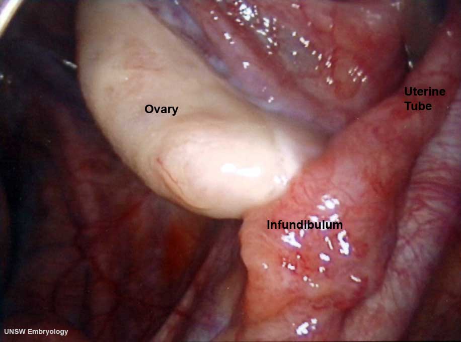
Human Ovary and Associated Uterine Tube
Adult human ovary (right) viewed by laparoscopy.
- Note the relative size and position of the ovary with respect to the uterine tube (fallopian tube, oviduct).
- The ovary surface appears white and relatively avascular, representing the dense connective tissue layer (tunica albuginea).
- In the background the associated mesenteries and peritoneal cavity can be seen.
- Links: ovary | uterus | Menstrual Cycle | Ovulation Image
Reference
UNSW Embryology
Copyright
No reproduction without permission.
Cite this page: Hill, M.A. (2024, June 5) Embryology Human right ovary and tube 1.jpg. Retrieved from https://embryology.med.unsw.edu.au/embryology/index.php/File:Human_right_ovary_and_tube_1.jpg
- © Dr Mark Hill 2024, UNSW Embryology ISBN: 978 0 7334 2609 4 - UNSW CRICOS Provider Code No. 00098G
File history
Click on a date/time to view the file as it appeared at that time.
| Date/Time | Thumbnail | Dimensions | User | Comment | |
|---|---|---|---|---|---|
| current | 09:31, 9 April 2010 | | 916 × 680 (32 KB) | S8600021 (talk | contribs) | Human right ovary and tube as viewed by laparoscopy == Image version links == Large 1000px | 800px | Medium 600px | [[:F |
You cannot overwrite this file.