File:Human CS13-15 otic vesicle 01.jpg

Original file (1,574 × 1,779 pixels, file size: 364 KB, MIME type: image/jpeg)
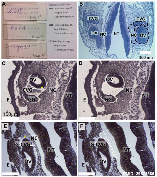
Laser-microdissection of FFPE-human inner ear slides. (A) FFPE slides containing embryonic tissues. (B) Coronal section of a fetus with highlighted regions of interest (dashed black circles, CVG, NC, and OV). Further images are provided showing tissue before (C, E) and after (D, F) laser-captured microdissection of CVG (dashed white lines in D) and NC tissues (dashed white line in F). Other developmental regions also are highlighted. CVG: cochlear-vestibular ganglions, OV: otic vesicle, NC: neural crest; GG: geniculate ganglion, G-CVG: geniculate- cochleovestibular ganglions, E: epithelium; and NT: neural tube. Scale bar = 100 μm (B) and 150 μm (C-F). https://doi.org/10.1371/journal.pone.0191452.g001
Reference
Chadly DM, Best J, Ran C, Bruska M, Woźniak W, Kempisty B, Schwartz M, LaFleur B, Kerns BJ, Kessler JA & Matsuoka AJ. (2018). Developmental profiling of microRNAs in the human embryonic inner ear. PLoS ONE , 13, e0191452. PMID: 29373586 DOI. Fig 1.
File history
Yi efo/eka'e gwa ebo wo le nyangagi wuncin ye kamina wunga tinya nan
| Gwalagizhi | Nyangagi | Dimensions | User | Comment | |
|---|---|---|---|---|---|
| current | 12:19, 6 April 2018 |  | 1,574 × 1,779 (364 KB) | Z8600021 (talk | contribs) | |
| 12:12, 6 April 2018 |  | 1,574 × 2,053 (457 KB) | Z8600021 (talk | contribs) | * '''Developmental profiling of microRNAs in the human embryonic inner ear'''{{#pmid:29373586|PMID29373586}} "Due to the extreme inaccessibility of fetal human inner ear tissue, defining of the microRNAs (miRNAs) that regulate development of the inner... |
You cannot overwrite this file.
File usage
The following page uses this file: