File:Anderson2016-fig10.jpg

Original file (800 × 800 pixels, file size: 109 KB, MIME type: image/jpeg)
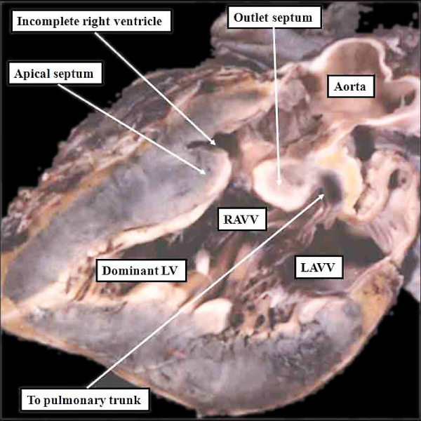
Fig. 10. Abnormal Heart
The image shows a congenitally malformed heart in which both the right and left atrioventricular valves (RAVV. LAVV) are connected with the dominant left ventricle. The right ventricle is incomplete, and is supplied through a ventricular septal defect, comparable to the embryonic interventricular communication shown in Figure 8, right hand panel. Note that, in this heart, the aorta arises from the incomplete right ventricle, and the pulmonary trunk from the dominant left ventricle. This is the arrangement usually described as “transposition”, but better accounted for in terms of discordant ventriculo-arterial connections.
File history
Yi efo/eka'e gwa ebo wo le nyangagi wuncin ye kamina wunga tinya nan
| Gwalagizhi | Nyangagi | Dimensions | User | Comment | |
|---|---|---|---|---|---|
| current | 14:56, 16 February 2017 | | 800 × 800 (109 KB) | Z8600021 (talk | contribs) | ==Fig. 10. Abnormal Heart== The image shows a congenitally malformed heart in which both the right and left atrioventricular valves (RAVV. LAVV) are connected with the dominant left ventricle. The right ventricle is incomplete, and is supplied through... |
You cannot overwrite this file.
File usage
The following 8 pages use this file:
- Cardiovascular System - Tricuspid Atresia
- Cardiovascular System - Ventricular Septal Defects
- Detailed Cardiac - Systemic Venous Sinus
- Lecture - Heart Development
- Paper - Teratogenecity in the setting of cardiac development and maldevelopment
- Template:Anderson2016 gallery1
- Template:Anderson2016 gallery2
- Template talk:Anderson2016 gallery1