File:MRI Human Embryo - upper limb 01.jpg
Original file (1,418 × 940 pixels, file size: 106 KB, MIME type: image/jpeg)
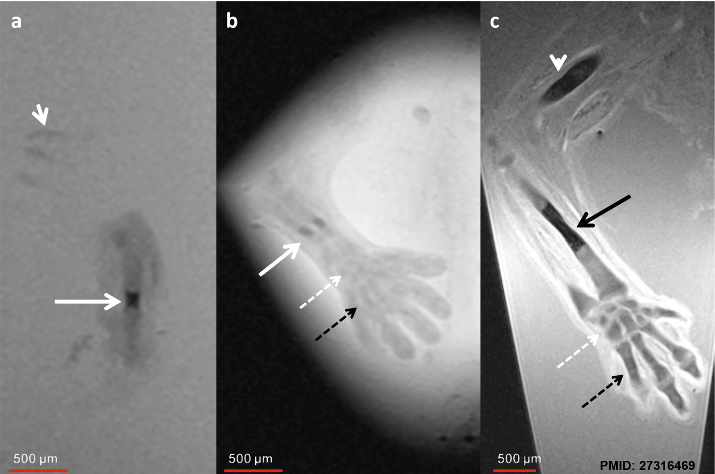
MRI Human Embryo - Upper Limb
Human embryo week 6 (GA 8 week) and week 7 (GA 9 week). Ex vivo magnetic resonance microscopy (MRM) at 7.1 T (Clin Scan, Bruker Biospin, Germany) was performed in 10 human specimens at 8 to 12 weeks of gestational age (GA). In-plane resolution was 20 μm with a slice thickness of 70 μm.
| a GA 8 week Sagittal T2w image of the humerus | b GA 8 week Coronal T2w image of the forearm | c GA 9 week Coronal T2w image |
| Shows initial ossification within the central part of the diaphysis (arrow). Chondrified ribs (short arrow). | width=300pxShows small ossification centers in the central parts of the radius and ulna (arrow). The carpal (dotted white arrow) and metacarpal bones (dotted black arrow) are already visible as precartilage states. | Shows increased size of the ossification centers in humerus (white arrowhead) and radius (black arrow). The carpal and metacarpal bones demonstrate progressive chondrification and appear hypointense compared to the 8-week GA specimen |
- Links: Magnetic Resonance Imaging
Reference
<pubmed>27316469</pubmed>
| width=300px|a Sagittal T2w image of the humerus of an 8-week GA specimen demonstrates initial ossification within the central part of the diaphysis (arrow). Chondrified ribs (short arrow).
| width=300px|b Coronal T2w image of the forearm of the same specimen as in a) demonstrates small ossification centers in the central parts of the radius and ulna (arrow). The carpal (dotted white arrow) and metacarpal bones (dotted black arrow) are already visible as precartilage states.
| width=300px|c Coronal T2w image of a 9-week GA specimen shows increased size of the ossification centers in humerus (white arrowhead) and radius (black arrow). The carpal and metacarpal bones demonstrate progressive chondrification and appear hypointense compared to the 8-week GA specimen
Copyright
© The Author(s). 2016
This article is distributed under the terms of the Creative Commons Attribution 4.0 International License (http://creativecommons.org/licenses/by/4.0/), which permits unrestricted use, distribution, and reproduction in any medium, provided you give appropriate credit to the original author(s) and the source, provide a link to the Creative Commons license, and indicate if changes were made. The Creative Commons Public Domain Dedication waiver (http://creativecommons.org/publicdomain/zero/1.0/) applies to the data made available in this article, unless otherwise stated.
Fig. 1 12861_2016_123_Fig1_HTML.gif
Cite this page: Hill, M.A. (2024, June 14) Embryology MRI Human Embryo - upper limb 01.jpg. Retrieved from https://embryology.med.unsw.edu.au/embryology/index.php/File:MRI_Human_Embryo_-_upper_limb_01.jpg
- © Dr Mark Hill 2024, UNSW Embryology ISBN: 978 0 7334 2609 4 - UNSW CRICOS Provider Code No. 00098G
File history
Click on a date/time to view the file as it appeared at that time.
| Date/Time | Thumbnail | Dimensions | User | Comment | |
|---|---|---|---|---|---|
| current | 22:50, 28 June 2016 | | 1,418 × 940 (106 KB) | Z8600021 (talk | contribs) | |
| 22:42, 28 June 2016 | 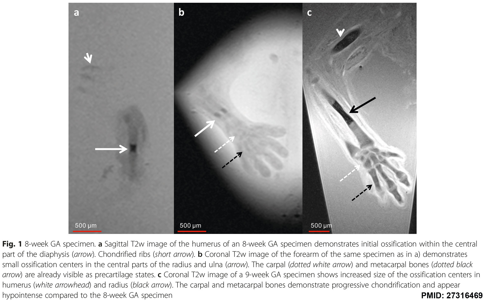 | 1,971 × 1,226 (258 KB) | Z8600021 (talk | contribs) | ==MRI Human Embryo - Upper Limb== Human embryo week 6 ({{GA}} 8 week). a Sagittal T2w image of the humerus of an 8-week GA specimen demonstrates initial ossification within the central part of the diaphysis (arrow). Chondrified ribs (short arrow). b... |
You cannot overwrite this file.
File usage
The following page uses this file: