Paper - The development of the cloaca in human embryos
| Embryology - 3 Jun 2024 |
|---|
| Google Translate - select your language from the list shown below (this will open a new external page) |
|
العربية | català | 中文 | 中國傳統的 | français | Deutsche | עִברִית | हिंदी | bahasa Indonesia | italiano | 日本語 | 한국어 | မြန်မာ | Pilipino | Polskie | português | ਪੰਜਾਬੀ ਦੇ | Română | русский | Español | Swahili | Svensk | ไทย | Türkçe | اردو | ייִדיש | Tiếng Việt These external translations are automated and may not be accurate. (More? About Translations) |
Pohlman AG. The development of the cloaca in human embryos. (1911) Amer. J Anat. 12: 1-26.
| Historic Disclaimer - information about historic embryology pages |
|---|
| Pages where the terms "Historic" (textbooks, papers, people, recommendations) appear on this site, and sections within pages where this disclaimer appears, indicate that the content and scientific understanding are specific to the time of publication. This means that while some scientific descriptions are still accurate, the terminology and interpretation of the developmental mechanisms reflect the understanding at the time of original publication and those of the preceding periods, these terms, interpretations and recommendations may not reflect our current scientific understanding. (More? Embryology History | Historic Embryology Papers) |
The Development of the Cloaca in Human Embryos
Augustus G. Pohlman
Indiana University
From the Anatomical Laboratory, Johns Hopkins University
Introduction
The active work in the embryology of the urogenital system began about 1880 and continued for a period of some fifteen years when Keibel's monograph was published. The writer's investigation of the cloacal region was started in 1903, and was undertaken in part as a control of Keibel's work, and in part with a view toward solving some of the points on which differences in opinion existed. The publication of this report has been delayed in the hope that certain facts in comparative embryology might be established and help to clarify some of the obscure relations found in the human embryo. Inasmuch as the extensive investigations of Fleischmann and his students have come to naught in this respect, the major differences in opinion will be considered, and the doubtful points answered in so far as it is possible. The short literature review covers the essential facts and effort has been made to reduce the description of the material to a concise tabulation. The writer expresses his indebtedness to Prof. Keibel at whose suggestion the development of the later stages in the embryology was undertaken, and to Prof. F. P. Mall for the use of his collection of embryos and for the many courtesies shown him in the Anatomical Laboratory of Johns Hopkins University.
Born's article ('93) reviews the earlier development of the cloacal region in a very complete manner, and the substance is as
follows: The entoderm of the enteron comes into direct relation
with the surface ectoderm in the pharyngeal and cloacal membranes
during the formation of the head and tail folds. Both of these
membranes lose their primitive position and become folded into
the substance of the embryo through increase in the surrounding
mesoderm. The allantois, which is developed dorsally in the
mammalian embryo (human and guinea pig excepted), shifts to a
ventral position on the gut, and is gradually displaced from its
intimate relation to the yolk sac through increase in the amount
of mesodermal tissue. The primitive streak is carried to the ventral surface of the body during the formation of the tail fold, and
forms the whole or part of the cloacal membrane. Kolliker
f'83), Strahl ('83, '84), and Bonnet ('88) believe that the caudal
end of the primitive streak is made up of applied layers of ectoand entoderm, and that it enters as such into the formation of the
cloacal membrane. Keibel ('88) argues that this primitive relation of the ecto- and entoderm is lost through interposition of
mesoderm; the latter disappearing later with restoration of the
original two layered condition.
The model of the 4.2 mm. human embryo presented by Keibel
('88) shows the hind gut and widely lumened allantois opening
cephalward into the caudal entodermal sac or cloaca. Ventrally,
this cloaca is limited as far as the dermal navel by the epithelial
cloacal membrane. Caudalward, the limit of the cloacal membrane comes about by a mesodermic separation of the epithelial
layers. The gut segment distal to the lower limit of the cloacal membrane may be termed the tail gut and terminates in an undifferentiated cell mass formed by itself, the chorda and neural tube. Born emphasizes the length of the cloacal membrane as follows: I call particular attention to the original extent of the cloacal membrane. It reaches cephalward to the point where the allantois leaves the body at right angles; i.e., as far as the caudal border of the dermal navel."
The cloacal membrane does not extend as far as the allantois
in the model of an 8.0 mm. human embryo presented by Keibel
('91) . Mesodermic tissue has apparently wandered in from above
and separated the layers of epithelium. The cloacal membrane
is therefore shorter than in the 4.2 mm. stage. The tail gut shows
degeneration. The precloacal mesodermic tissue has increased
in amount but instead of displacing the cloacal membrane caudal-ward, has folded it into the lower surface of the genital tubercle
as was first described by Tourneux ('89), and verified by Retterer
('90) and Reichel ('93). No intermediate stage in the development has been described up to this time ('93). With the increase
in size of the genital tubercle, the entodermal cloaca becomes
more deeply placed as was demonstrated by Reichel ('93). Born
states that the epithelial plate contained within the genital tubercle is ectodermic and that it is continuous with the superficial
ectoderm. The epithelial plate occupies the caudal surface of
the eminence and is bordered laterally by folds of mesoderm (repli
ano-genitaux of Retterer) , while the cloacal membrane itself terminates at the postanal fold (replis postanal)., The depression arises (as in the mouth region) through increase in the height of the limiting borders. The depression is
always closed in by epithelium, and the base of the depression is
never separated from the entodermal cloaca by mesodermic tissue." (Born.)
The cloaca is gradually divided into a ventral (bladder-urogenital sinus) segment, and a dorsal (gut) segment. As to the
manner of this division, Retterer and Born agree with the Tiedemann-Rathke idea of the gradual separation into two segments
through approximation of two lateral folds of mesoderm, while Tourneux believes it to be accomplished by a septal (frontal) downgrowth. Lieberkiihn ('82) and Keibel ('89) dispute the
theory of Rathkc ('32) championed bj' von Mihlacovics ('85),
that the bladder arises from the allantois, and state that it is made
up for the most part from the ventral cloacal segment — agreed
to by Retterer and Reichel. Born takes a neutral position and
believes that at least the trigone of the human bladder may be
developed in a manner like that found in the guinea pig (Keibel) .
Born and Minot do not think that the anlage of the upper part
of the bladder is of particular importance. "We are probably
not mistaken when we grant that not only the bladder (as far as the apex) but the male urethra as far as the caput gallinaginis, the entire female urethra, and in the male, also the pars prostatica and the entire pars membranacea are developed from the ventral cloacal segment."
The most important of the recent works is that of Ke bel ('96) who sums up the development of the cloacal region in the following words : The human embryo possesses a large entodermal cloaca in the early stages of its development which however is never opened to the outside through a cloacal anus ('Cloakenafter' of Prenant), but remains closed through the cloacal membrane (the 'anal membrane' of the earlier writers) as long as it exists as such. An ectodermal cloaca is to be found only in traces if at
all. The entodermal cloaca is separated into a ventral and a dorsal segment by a frontal septum. A large part or all of the bladder,
the urethra and the urogenital sinus as far as the cloacal membrane
are derived from the ventral segment; while the dorsal segment
beconjes continuous with the ectodermal segment of the rectal
canal. The primitive perineum is formed when the frontal septum fuses with the cloacal membrane and the rudimentary ectodermal cloaca is then divided by the permanent perineum. The
ectodermal anal pit (protodaeum) is situated behind the permanent perineum, while the ectodermal portion of the urogenital sinus is ventral to it."
Another investigator, who has been particularly fortunate in
the amount of embryological material at his disposal, expresses
a somewhat different view. Nagel ('02) practicall}- reiterates
his statements of 1894. "The inspection of the tail end of human embryos of 11-13 mm. length reveals an oval pit extending from
the coccygeal prominence to the tip of the genital eminence.
This pit (cloaca) receives the openings of the gut dorsally, and the
Canalis urogenitalis ventrally; the two separated by a partition
of some 0.3 mm. thickness. The Wolffian and Miillerian ducts
open higher up in the Canalis urogenitalis and will not be considered in the description of this depression. The Canalis urogenitalis and the gut open into this pit (cloaca) which would reach
(comparing with adult relations) from the dorsal border of the
anus to the ventral border of the urethral opening {i.e. Frenulum
clitoridis). Later he states: In what manner the division of
the cloaca is accomplished is not perfectly understood either in
man or in mammals. I found the relations in the youngest human embryo that I had opportunity to examine like those pictured
in fig, 1, naturally with exception of the form of the bladder.
I commit myself therefore, as far as the human embryo is concerned, to the view of Rathke which has recently been substantiated by Retterer and von Mihalcovics in the animals."
Fig. 1
The work of von Mihalcovics i/85) referred to, says in part; This septum between the urogenita' canal and the gut arises in part through increase in length of the afore-mentioned perineal Told (septum) to which, distalward, two lateral folds join to the perineal folds. The cloaca takes no part in the formation of the urogenital canal. "The opinion of Retterer ('94), mentioned by Nagel, is summarized as follows: "In the guinea pig, as in other mammals studied up to the present time (man, pig, sheep and rabbit) , a fold of mesoderm appears at the cephalic extremity of each lateral cloacal wall, and extends little by little toward the caudal end of the cloaca. These lateral folds encroach upon the lumen of the cloaca and divide it into two canals, the one dorsal or rectal, and the other ventral or vesico-urogenital."
That these opinions, while they are in the main contrary to the views of Keibel although they appeared at an earlier date, are accepted at the present time can be illustrated by an extract from Zuckerakndls Handbuch der Urologie ('03): In the second fetal month, the proximal segment of the allantois widens to form the bladder, while the distal and narrow portion (urachus) obliterates to form the Lig. vesico-umbilicale, "The division of the cloaca is accomplished by three folds, a median and two lateral. The former occupies the angle between the allantois and hind gut, while the latter are developed in the lateral walls of the cloaca."
The investigations of Tourneux ('94) agree with those of Keibel
in that he also states the cloaca to be a closed sac. His objections to the method of cloacal division as described byRetterer
are as follows: The form of the inferior border of the recto-urogenital septum is that of a vaulted arch and not that of an
elliptical arch with thevertex upward — afact easity demonstrated
in frontal sections. In addition to this, the transverse sections
show that the lateral folds are found only toward the summit of the
arch and converge rapidly. Further, the septum shows no signs
of an epithelial raphe at the supposed line of fusion to indicate
the transition that one encounters, as Keibel states, at the line
of union of the palatine ridges."
Nagel ('96) answers Keibel's article by stating If Keibel did
not find the facts as presented by myself in all of his embryos of
11.0 mm. and upwards, then the embryos are at fault." "Furthermore in order that an embryo may be declared of scientific
value, I demand that the urogenital canal be open into the cloacal
pit in all embryos over 8.0 mm., and that the cloacal membrane
have disappeared as far as the tip of the genital eminence. Inasmuch as th& allantois contained within the umbilical cord is practically obliterated at this stage, where could the secretion from the
mesonephros be stored up if the cloacal membrane were intact?"
The questions that appeal to the writer as doubtful ones may be expressed as follows:
- Does the cloaca proper ever open to the outside?
- Does the cloacal membrane arise directly by displacement of applied layers of ecto- and entoderm in the primitive streak or is it formed in situ through disappearance of the intervening mesoderm?
- In what manner is the cloacal division effected?
- What is the anlage for the bladder?
- Can the urinary function of the mesonephros assumed by Nagel be demonstrated?
Material
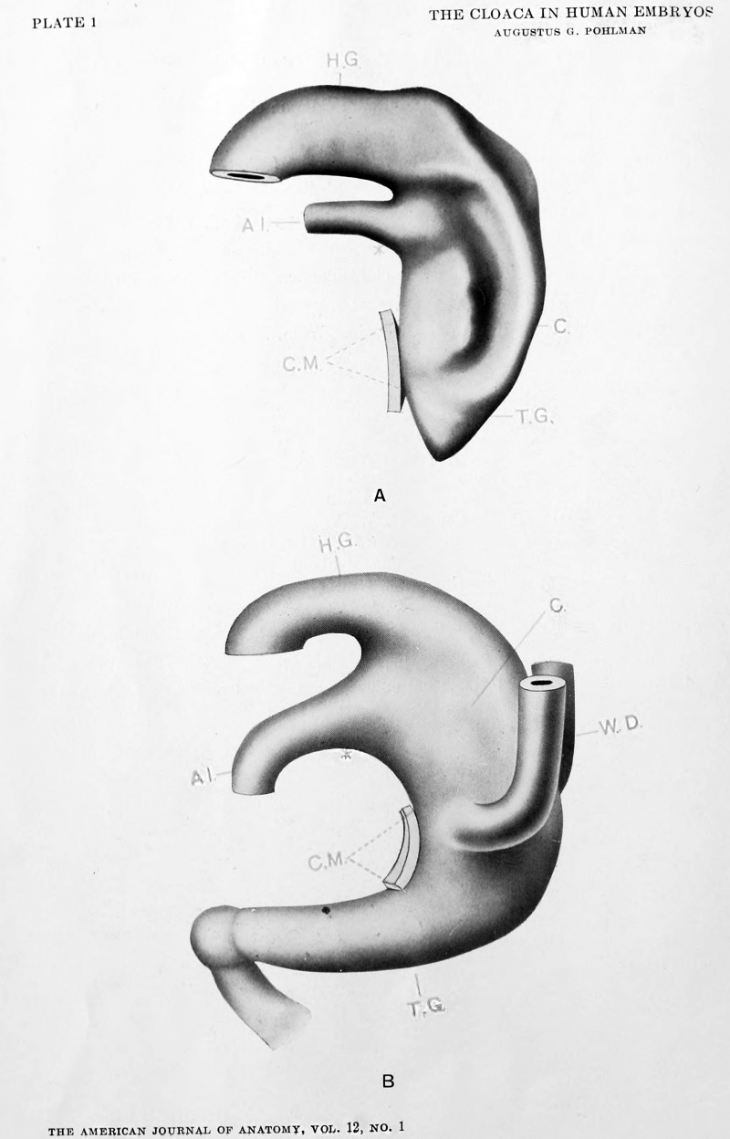
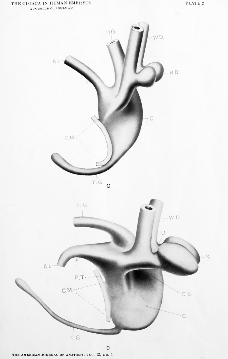
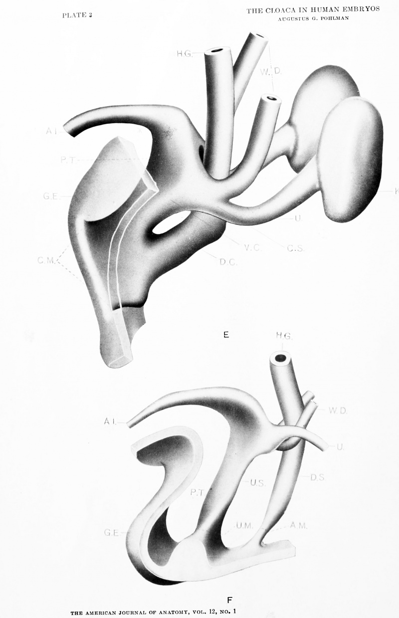
The study of the cloacal region was done in part by working out the relations in serial sections, and in part through reconstruction. Thirty embryos in all were examined; reconstruction employed in thirteen, of which six stages will be presented. The material, with the exception of these six , is given entirely in the tabulation. The serial number, used throughout this paper, refers to the age of the embryo based on the development of the urogenital tract and with no particular reference to its length. It is interesting to note that with the exception of nos. 4, 11, 13, and 21, the development of the tract has proved to be an excellent check on the determination of the age by the greatest length method. The Mall number refers to the catalogue number of the collection of human embryos at Johns Hopkins University and the section thickness (indicated in microns) ; the section direction (+ for transverse, = for sagittal, and || for coronal; and the condition (/ for fair and g for good) are recorded in this manner in the Mall catalogue. The modelling of the embryo is indicated by the magnification of the same, and where the model is presented in this paper it is designated by a capital letter. The brief normentafel shows the major points of difference in the development of the cloacal region. The embryo 30 is the Piper II embryo of the collection, and will be presented later in connection with the origin of the bulbovestibular glands. This embryo and also no. 20 have been reported in connection with the condition of complete double ureter (Johns Hopkins Bull., vol. 16, Feb. '04).
Tabulation
Tabulation of Material. + for transverse: = for sagittal: || for coronal. The figures following indicate the thickness in microns.
No. 1 (Mall 12; 2.1 mm.; +; 10; Good.) Modelled by Mall ('97). Allantois of small lumen and in relation with the caudal border of the yolk sac. No evidences of a cloacal membrane.
No. 2 (Mall 391 ; 2.0 mm. ; + ; 10; Good). Appearance of hind gut. An interval of several sections between allantois and yolk sac. No cloacal membrane.
No. 3 (Mall 164; 3.5 mm. ; + ; 20; Good). Model A. X 100. Cloaca well developed and limited for caudal half of the ventral border by epithelial cloacal membrane. Allantois comes off at right angles and at lower border of dermal navel. Lateral furrow along later line of division.
No. 4 (Mall 209; 3.0 mm. ; + ; 50; Fair). Relations about the same as the fore-going stage. Sections too thick for detailed study.
No. 5 (Mall 186; 3.5 mm.; +; 20; Good). Model B. X 100. Allantois wide lumened and no distinct line of demarkation from the ventral cloacal segment. Wolflian ducts have arrived but not as yet opened. Tail gut maximum of development. Probably the first signs of cloacal division.
No. 6 (Mall 87; 4.0 mm.; +; 20; Fair). About the same as the foregoing.
No. 7 (Mall 148; 4.3 mm. ; + ; 10; Good X 100). Wolffian ducts open into cloaca. Allantois of small lumen. Ventral cloacal segment widening. Evidences of division quite apparent.
No. 8 (Mall. 76; 4.5 mm. ; + ; 20; Good). X 100. Widened ventral cloacal segment goes over indistinctly into allantois. Tail gut still quite large. Renal buds well developed. With exception of large allantois and wider tail gut, quite similar to Model C.
No. 9 (Mall 80; 5.0 mm; + ; 10; Good). Model C. X 100. About the same as the foregoing stage. Tail gut undergoing degenerative changes.
No. 10 (Mall 371; 6.6 mm.; = ; 10; Good). Tail gut rudimentary. Development of cloaca and renal buds about the same as in no. 12.
No. 11 (Mall 116; 5.0 mm. ; = ; 20; Good). X 100. Intracloacal epithelial plug well marked. Stage about the same as that found in no. 12.
No. 12 (Mall 2; 7.0 mm.; +; 15; Good). Model D. X 100. Marked development of renal buds. Kidney and ureter segments evident. Ventral cloacal segment much widened. Allantois of narrow lumen. Intracloacal epithelial plug marked. Tail gut maximum in length and slightly widened at caudal end.
No. 13 (Mair241;6.0mm.; +; 10;Goo(l). Tailgutmore rudimentary than in no. 12. Cloacal membrane wide and shows intracloacal epithelial plug. Otherwise about the same as in no. 12.
No. 14 (Mall 383; 7.0 mm.; +," 10; Good). State similar to no. 12. Cloacal membrane intact.
No. 15 (Mall 397; 8.0 nma.; +; 10; Good). Similar to foregoing.
Plates
Cite this page: Hill, M.A. (2024, June 3) Embryology Paper - The development of the cloaca in human embryos. Retrieved from https://embryology.med.unsw.edu.au/embryology/index.php/Paper_-_The_development_of_the_cloaca_in_human_embryos
- © Dr Mark Hill 2024, UNSW Embryology ISBN: 978 0 7334 2609 4 - UNSW CRICOS Provider Code No. 00098G


