Paper - The development of the cloaca in human embryos
| Embryology - 3 Jun 2024 |
|---|
| Google Translate - select your language from the list shown below (this will open a new external page) |
|
العربية | català | 中文 | 中國傳統的 | français | Deutsche | עִברִית | हिंदी | bahasa Indonesia | italiano | 日本語 | 한국어 | မြန်မာ | Pilipino | Polskie | português | ਪੰਜਾਬੀ ਦੇ | Română | русский | Español | Swahili | Svensk | ไทย | Türkçe | اردو | ייִדיש | Tiếng Việt These external translations are automated and may not be accurate. (More? About Translations) |
Pohlman AG. The development of the cloaca in human embryos. (1911) Amer. J Anat. 12: 1-26.
| Historic Disclaimer - information about historic embryology pages |
|---|
| Pages where the terms "Historic" (textbooks, papers, people, recommendations) appear on this site, and sections within pages where this disclaimer appears, indicate that the content and scientific understanding are specific to the time of publication. This means that while some scientific descriptions are still accurate, the terminology and interpretation of the developmental mechanisms reflect the understanding at the time of original publication and those of the preceding periods, these terms, interpretations and recommendations may not reflect our current scientific understanding. (More? Embryology History | Historic Embryology Papers) |
The Development of the Cloaca in Human Embryos
Augustus G. Pohlman
Indiana University
From the Anatomical Laboratory, Johns Hopkins University
Introduction
The active work in the embryology of the urogenital system began about 1880 and continued for a period of some fifteen years when Keibel's monograph was published. The writer's investigation of the cloacal region was started in 1903, and was undertaken in part as a control of Keibel's work, and in part with a view toward solving some of the points on which differences in opinion existed. The publication of this report has been delayed in the hope that certain facts in comparative embryology might be established and help to clarify some of the obscure relations found in the human embryo. Inasmuch as the extensive investigations of Fleischmann and his students have come to naught in this respect, the major differences in opinion will be considered, and the doubtful points answered in so far as it is possible. The short literature review covers the essential facts and effort has been made to reduce the description of the material to a concise tabulation. The writer expresses his indebtedness to Prof. Keibel at whose suggestion the development of the later stages in the embryology was undertaken, and to Prof. F. P. Mall for the use of his collection of embryos and for the many courtesies shown him in the Anatomical Laboratory of Johns Hopkins University.
Born's article ('93) reviews the earlier development of the cloacal region in a very complete manner, and the substance is as
follows: The entoderm of the enteron comes into direct relation
with the surface ectoderm in the pharyngeal and cloacal membranes
during the formation of the head and tail folds. Both of these
membranes lose their primitive position and become folded into
the substance of the embryo through increase in the surrounding
mesoderm. The allantois, which is developed dorsally in the
mammalian embryo (human and guinea pig excepted), shifts to a
ventral position on the gut, and is gradually displaced from its
intimate relation to the yolk sac through increase in the amount
of mesodermal tissue. The primitive streak is carried to the ventral surface of the body during the formation of the tail fold, and
forms the whole or part of the cloacal membrane. Kolliker
f'83), Strahl ('83, '84), and Bonnet ('88) believe that the caudal
end of the primitive streak is made up of applied layers of ectoand entoderm, and that it enters as such into the formation of the
cloacal membrane. Keibel ('88) argues that this primitive relation of the ecto- and entoderm is lost through interposition of
mesoderm; the latter disappearing later with restoration of the
original two layered condition.
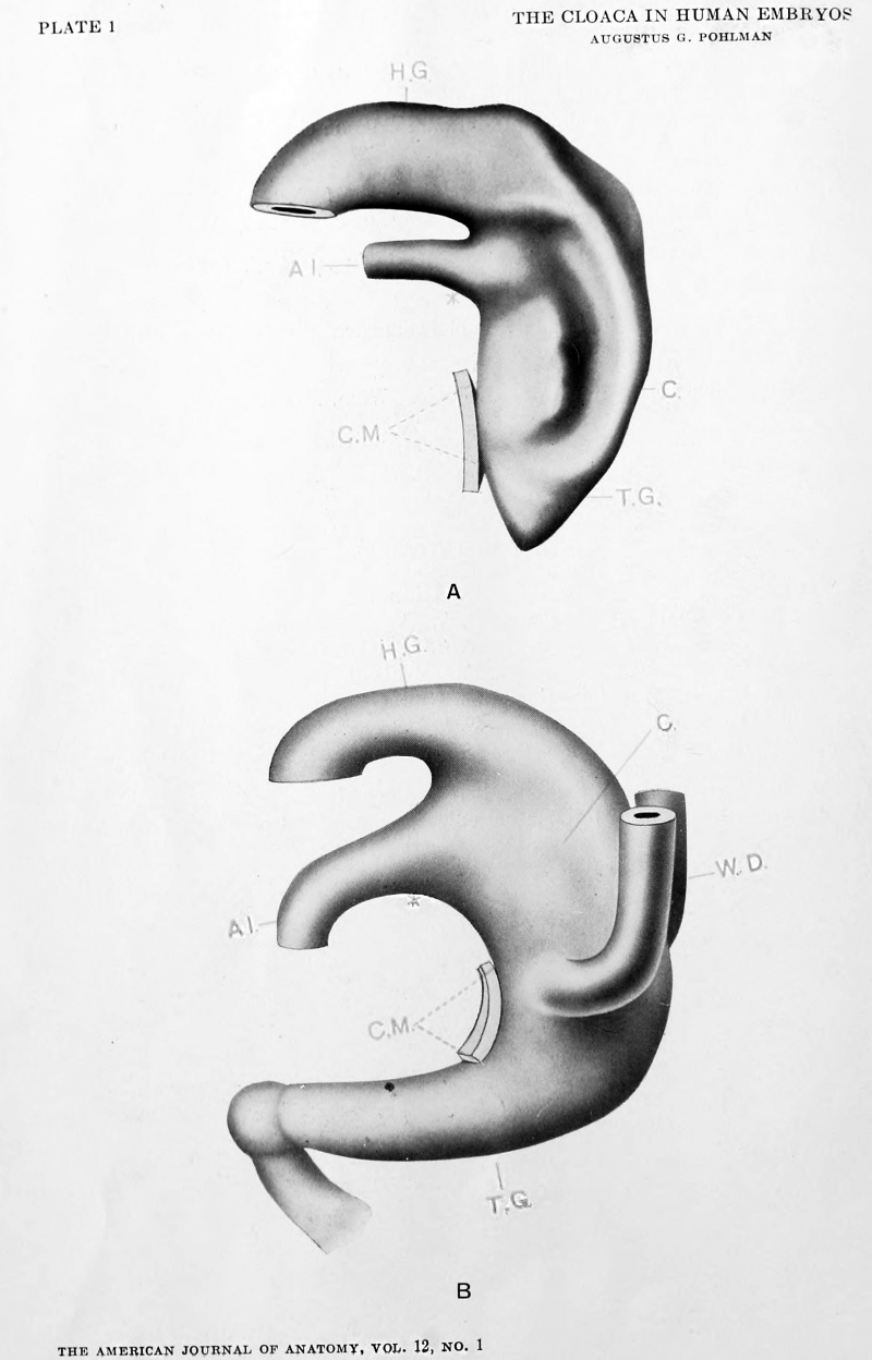
The model of the 4.2 mm. human embryo presented by Keibel
('88) shows the hind gut and widely lumened allantois opening
cephalward into the caudal entodermal sac or cloaca. Ventrally,
this cloaca is limited as far as the dermal navel by the epithelial
cloacal membrane. Caudalward, the limit of the cloacal membrane comes about by a mesodermic separation of the epithelial
layers. The gut segment distal to the lower limit of the cloacal
Cite this page: Hill, M.A. (2024, June 3) Embryology Paper - The development of the cloaca in human embryos. Retrieved from https://embryology.med.unsw.edu.au/embryology/index.php/Paper_-_The_development_of_the_cloaca_in_human_embryos
- © Dr Mark Hill 2024, UNSW Embryology ISBN: 978 0 7334 2609 4 - UNSW CRICOS Provider Code No. 00098G


