File:Human week 10 fetus 10.jpg
From Embryology
Size of this preview: 800 × 600 pixels. Other resolution: 1,200 × 900 pixels.
Original file (1,200 × 900 pixels, file size: 291 KB, MIME type: image/jpeg)
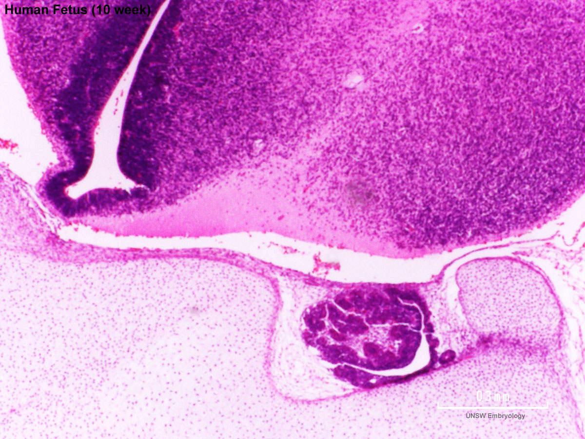
Human Female Fetus - Pituitary and Lamina Terminalis (10 week)
Large image version of plane D, close to midline (Stain - Haematoxylin Eosin) 0.3 mm scale bar
- Lamina Terminalis - site of anterior neuropore closure.
- Pituitary - anterior pituitary is shown in this section, lying within the sella turcica.
- Hypothalamus - brain region shown above the pituitary.
- Sphenoid - still cartilage at this stage, the base of the skull ossifies by endochondral ossification.
- Human Female Fetus (week 10)
Sagittal Section (plane D)

Pituitary and Lamina Terminalis

Olfactory Nerve

Atlas and Axis

Sacrum

Oral Cavity

Epiglottis

Heart

Spleen

Midgut Herniation

Midgut Herniation (label)

Pelvic Region

Pelvic Region (label)


Related Images
Fetus (week 10) Planes A (most lateral), B (lateral), C (medial) and D (midline) from lateral towards the midline.
- Human Fetus - most lateral | lateral | medial | midline
- Head - most lateral | lateral | medial | midline
- Cerebellum - most lateral | lateral | medial | midline
- Urogenital Unlabelled - most lateral | lateral | medial | midline
- Urogenital Labelled - most lateral | lateral | medial | midline
- Large Images - midline
- Image Source: UNSW Embryology, no reproduction without permission.
File history
Click on a date/time to view the file as it appeared at that time.
| Date/Time | Thumbnail | Dimensions | User | Comment | |
|---|---|---|---|---|---|
| current | 23:05, 17 June 2012 | | 1,200 × 900 (291 KB) | Z8600021 (talk | contribs) | ==Human Female Fetus Pituitary (10 week)== Large image version of plane D, close to midline (H&E stain). 0.5 mm scale bar Note: {{10wkFetus}} |
You cannot overwrite this file.
File usage
The following 20 pages use this file:
- BGDA Practical 12 - Embryo to Fetus
- BGDB Gastrointestinal - Fetal
- Fetal Development - 10 Weeks
- Foundations Practical - Week 9 to 36
- L
- Neural System Development
- File:Human week 10 fetus 01.jpg
- File:Human week 10 fetus 03.jpg
- File:Human week 10 fetus 04.jpg
- File:Human week 10 fetus 05.jpg
- File:Human week 10 fetus 06.jpg
- File:Human week 10 fetus 07.jpg
- File:Human week 10 fetus 08.jpg
- File:Human week 10 fetus 09.jpg
- File:Human week 10 fetus 10.jpg
- File:Human week 10 fetus 11.jpg
- File:Human week 10 fetus 12.jpg
- File:Human week 10 fetus 23.jpg
- File:Human week 10 fetus 26.jpg
- Template:Human Female Fetus Week 10 gallery