File:Human embryonic-fetal tongue 01.jpg

Original file (1,000 × 1,129 pixels, file size: 490 KB, MIME type: image/jpeg)
Organogenesis of a fetal tongue. Photographs of mid-sagittal sections of an embryonic human tongue. (A, B) Tongue development (TD) stage 1. Co, copula; Ce, foramen cecum. (C) TD stage 1. (D, E) TD stage 2. (F, G) TD stage 3. (H) TD stage 4. (I, J) TD stage 5. MC, Meckerl's cartilage; Md, mandible. (K) TD stage 6. (L) TD stage 7. Microscopic features of tongue muscle at TD stage 3 (M), TD stage 6 (N), and TD stage 7 (O).
Tongue Growth during Prenatal Development in Korean Fetuses and Embryos
Soo Jeong Hong, Bong Geun Cha1, Yeon Sook Kim2, Suk Keun Lee, Je Geun Chi3 Department of Oral Pathology, College of Dentistry, Gangnueng-Wonju National University, Gangneung, Korea 1Department of Orthodontics, College of Dentistry, Gangnueng-Wonju National University, Gangneung, Korea 2Department of Dental Hygiene, College of Health Sciences, Cheongju University, Cheongju, Korea 3Department of Pathology, Seoul National University College of Medicine, Seoul, Korea Corresponding Author: Suk Keun Lee, DDS Department of Oral Pathology, College of Dentistry, Gangnueng-Wonju National University, 7, Jukheon-gil, Gangneung 25457, Korea Tel: +82-33-640-2228 Fax: +82-33-642-6410 E-mail:sukkeunlee@hanmail.net Received September 13, 2015 Accepted September 17, 2015 © 2015 The Korean Society of Pathologists/The Korean Society for Cytopathology This is an Open Access article distributed under the terms of the Creative Commons Attribution Non-Commercial License (http://creativecommons.org/licenses/by-nc/3.0/) which permits unrestricted non-commercial use, distribution, and reproduction in any medium, provided the original work is properly cited.
Fig. 1. Jptm-2015-09-17f1.jpg
File history
Yi efo/eka'e gwa ebo wo le nyangagi wuncin ye kamina wunga tinya nan
| Gwalagizhi | Nyangagi | Dimensions | User | Comment | |
|---|---|---|---|---|---|
| current | 13:56, 23 March 2016 | | 1,000 × 1,129 (490 KB) | Z8600021 (talk | contribs) | |
| 13:41, 23 March 2016 | 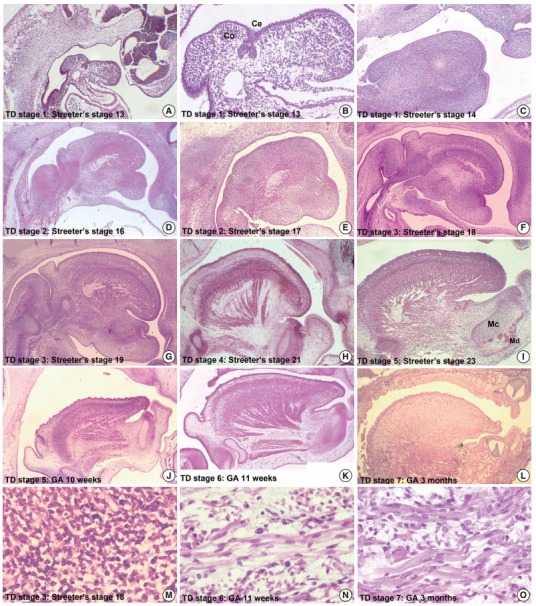 | 536 × 606 (147 KB) | Z8600021 (talk | contribs) | Organogenesis of a fetal tongue. Photographs of mid-sagittal sections of an embryonic human tongue. (A, B) Tongue development (TD) stage 1. Co, copula; Ce, foramen cecum. (C) TD stage 1. (D, E) TD stage 2. (F, G) TD stage 3. (H) TD stage 4. (I, J) TD s... |
You cannot overwrite this file.
File usage
The following page uses this file:


