Talk:Draft 2016

From Embryology

Lymph Nodes

Lymph Node Anatomy

Lymph Node Anatomy
  • Location throughout the entire body - concentrated in axilla, groin, lung, gastrointestinal tract mesenteries
  • Small (1 mm - 2 cm) encapsulated organ (diffuse lymphoid tissue - no capsule)
  • Antigen transformed lymphocytes from the blood
  • In lymph vessel pathways “filter” lymph
    • Afferent - towards node (A - arrives at the node)
    • Efferent - away from node (E - exits the node)

Gastrointestinal tract intestine immune cartoon 01.jpg

Mesenteric lymph nodes

Lymphatic-system-overview.jpg

Lymph Node Functions

Lymph Node Functions 
  • In lymph vessel pathways “filter” (surveillance) lymph
  • Immune - detect infections from peripheral tissues
    • skin, respiratory tract, gastrointestinal tract, etc.
  • secondary lymphoid organ
  • return extracellular fluid to circulation
Lymph node structure.jpg

Lymph Node Structure

Lymph Node Structure 
Lymph node cartoon.jpg

Simplified structure

Lymph node cartoon 02.jpg

Lymphocyte (T and B) Traffic

  1. Enter from high endothelial venules (HEVs also called post-capillary venules)
  2. Spend 8 to 24 h in the lymph node interstitium.
  3. Enter a network of medullary sinuses.
  4. Drain from sinuses into efferent lymphatic vessels.
Lymph node structure 02.jpg Lymph pathway
  1. Afferent vessel
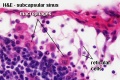
  2. Subcapsular sinus
  3. Paratrabecular sinus
  4. Medullary sinus
  5. Efferent vessel

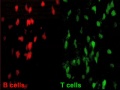
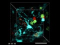
Watch T and B Lymphocytes Move 

Mouse adult lymph node 07.jpg
 ‎‎Lymph Node 7
Page | Play
Mouse adult lymph node 08.jpg
 ‎‎Lymph Node 8
Page | Play
Lymph node cartoon 01.jpg

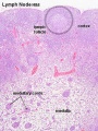
Lymph Node Histology

Lymph Node Histology 
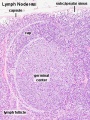
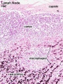
  • Capsule (thin) with trabecular or septa (dense connective tissue)

Lymph node cartoon 03.jpg


Fetal thymus Young medulla Young cortex
Fetal thymus.jpg Thymus - young 01.jpg Thymus - young 02.jpg


Lymph Node Cartoons: Detailed structure | Cartoon with Histology | Lymphocyte traffic | Simple structure | Simple node anatomy | Wiki node image | Internal structure | Mesenteric lymph node | Histology | Gallery | Lymph Node Development

Lymph Nodes

Lymph Node Structure

Lymph_node_cartoon_01.jpg

Connective Tissue

  • Capsule - dense connective tissue (irregular CT, some adipocytes))
  • Trabeculae - dense connective tissue
  • Reticular Tissue - Reticular cells and fibers, supporting meshwork (collagen type III)
    • Reticular cell produces reticular fibers (collagen type III) and surrounds the fibers with its cytoplasm
    • reticular fibbers can also be produced by fibroblasts
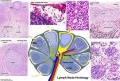

Lymphocyte (T and B) Traffic

  1. Enter from high endothelial venules (HEVs also called post-capillary venules)
  2. Spend 8 to 24 h in the lymph node interstitium.
  3. Enter a network of medullary sinuses.
  4. Drain from sinuses into efferent lymphatic vessels.


Important - Please note that the lymphocytes exit from the node by efferent lymphatic vessels.

See Figure

See also Image - Cell Trafficking into and out of Lymph Nodes.

Mouse adult lymph node 07.jpg
 ‎‎Lymph Node 7
Page | Play

T and B motility

Mouse adult lymph node 08.jpg
 ‎‎Lymph Node 8
Page | Play

T and B interaction

Lymph node histology 05.jpg

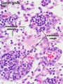
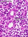
High Endothelial Venules

See also Lymphocyte Migration at High Endothelial Venule Model

Links: Immunobiology - Figure 1.8. Organization of a lymph node