Category:Pharyngeal Arch
From Embryology
This Embryology category covers content related to pharyngeal (branchial) arch development. This is generally covered in Head Development notes.
Pages in category 'Pharyngeal Arch'
The following 54 pages are in this category, out of 54 total.
C
P
- Paper - Evolutionary factors in the production of pharyngeal diverticula
- Paper - On the relation of the head chorda to the pharyngeal epithelium in the pig embryo
- Paper - The aortic arch derivatives in human adult (1951)
- Paper - The development of the first branchial arch in man and the fate of Meckel's cartilage
- Paper - The Disappearance of the Precervical Sinus
- Paper - The fifth aortic arch of mammalian embryos; the nature of the last pharyngeal evagination
- Paper - The pharyngeal pouches and their derivatives in the mammalia
- Paper - The potency of the pharyngeal entoderm (1932)
- Paper - The second visceral arch and groove in the tubo-tympanic region
- Paper - Three demonstrations on congenital melformations of palate, face, and neck
- Paper - Transformation of the aortic-arch system during the development of the human embryo (1922)
- Template:Pharyngeal arch
- Template:Pharyngeal Arch collapse table
- Template:Pharyngeal Arch table
- Pharyngeal arches
R
- Template:Ref-Allis1923
- Template:Ref-Anderson1922
- Template:Ref-Barry1951
- Template:Ref-Coulter1909
- Template:Ref-Frazer1914
- Template:Ref-Frazer1926
- Template:Ref-Keith1909
- Template:Ref-Kingsbury1914b
- Template:Ref-Negus1925
- Template:Ref-Rand1917
- Template:Ref-Reagan1912
- Template:Ref-Shaner1921
- Template:Ref-Woollard1932
- Template:Reichert’s cartilage
Media in category 'Pharyngeal Arch'
The following 51 files are in this category, out of 51 total.
 Congdon1922-31.jpg 1,063 × 1,000; 93 KB
 Congdon1922-32.jpg 1,133 × 1,000; 132 KB
 Congdon1922-34.jpg 920 × 1,000; 122 KB
 Congdon1922-35.jpg 920 × 1,000; 97 KB
 Congdon1922-36.jpg 920 × 1,000; 113 KB
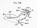 Foster136.jpg 556 × 420; 33 KB
 Gray0947.jpg 600 × 398; 56 KB
 Gray0978.jpg 483 × 600; 67 KB
 Gray0979.jpg 500 × 446; 56 KB
 Gray0980.jpg 542 × 450; 57 KB
 Gray0981.jpg 538 × 340; 50 KB
 Head and heart muscle cartoon.jpg 874 × 800; 129 KB
 Heart outflow tract stage 14 03.jpg 989 × 996; 134 KB
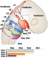 Hindbrain neural crest migration.jpg 450 × 545; 48 KB
 Human stage16 face 01.jpg 500 × 504; 20 KB
 Human stage17 face 01.jpg 500 × 504; 21 KB
 Human stage18 face 01.jpg 500 × 504; 23 KB
 Keibel Mall 2 314.jpg 760 × 1,000; 103 KB
 Keibel Mall 2 315.jpg 604 × 800; 61 KB
 Keibel Mall 2 316.jpg 899 × 1,000; 148 KB
 Keibel Mall 2 317.jpg 805 × 1,000; 95 KB
 Keibel Mall 2 318.jpg 1,077 × 1,000; 111 KB
 Keibel Mall 2 319.jpg 811 × 800; 82 KB
 Keibel Mall 2 320.jpg 973 × 800; 110 KB
 Keibel Mall 2 321.jpg 904 × 800; 76 KB
 Keibel Mall 2 322.jpg 729 × 800; 47 KB
 Keibel Mall 2 323.jpg 718 × 800; 54 KB
 Keibel Mall 2 324.jpg 1,280 × 930; 125 KB
 Keibel Mall 2 325.jpg 1,280 × 882; 225 KB
 Keibel Mall 2 326.jpg 1,280 × 730; 207 KB
 Keibel Mall 2 327.jpg 1,100 × 676; 114 KB
 Keibel Mall 2 328.jpg 1,100 × 534; 96 KB
 Keibel Mall 2 329.jpg 930 × 582; 74 KB
 Keibel Mall 2 330.jpg 1,280 × 1,009; 342 KB
 Meckels cartilage - middle ear from the jaw.jpg 1,161 × 1,280; 259 KB
 Pharyngeal arch cartilages.jpg 400 × 324; 26 KB
 Stage 13 image 005.jpg 1,000 × 451; 81 KB
 Stage 13 image 006.jpg 1,000 × 439; 83 KB
 Stage 13 image 007.jpg 1,000 × 514; 93 KB
 Stage 13 image 056.jpg 1,000 × 516; 102 KB
 Stage 13 image 057.jpg 1,000 × 511; 99 KB
 Stage 13 image 058.jpg 1,000 × 481; 94 KB
 Stage 13 image 059.jpg 1,000 × 513; 92 KB
 Stage 13 image 060.jpg 1,000 × 486; 96 KB
 Stage 13 image 061.jpg 1,000 × 600; 101 KB
 Stage13 face ventral view01.jpg 1,290 × 2,048; 152 KB
 Stage13 oral cavity floor01.jpg 1,315 × 2,048; 234 KB
 Stage13 oral cavity floor02.jpg 1,315 × 2,048; 371 KB
 Stage13 pharyngeal arch excerpts.gif 600 × 300; 86 KB
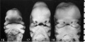 Stage16-18 face.jpg 800 × 393; 34 KB
 West02.jpg 619 × 549; 34 KB


