File:Human brain growth 01.jpg
From Embryology
Size of this preview: 766 × 600 pixels. Other resolution: 1,022 × 800 pixels.
Original file (1,022 × 800 pixels, file size: 119 KB, MIME type: image/jpeg)
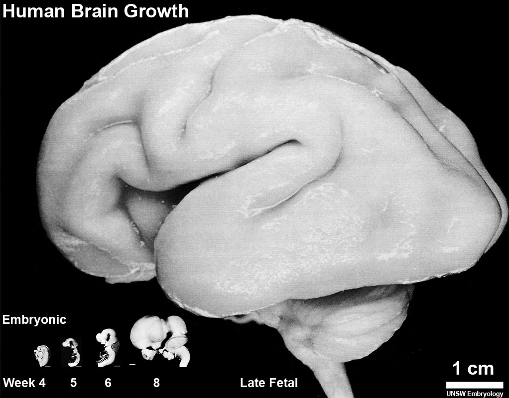
Human Brain Growth
Left lateral view of the relative overall size of the developing human brain during the embryonic period (week 4, 5, 6, and 8) and late fetal (third trimester close to term).
Cite this page: Hill, M.A. (2024, June 26) Embryology Human brain growth 01.jpg. Retrieved from https://embryology.med.unsw.edu.au/embryology/index.php/File:Human_brain_growth_01.jpg
- © Dr Mark Hill 2024, UNSW Embryology ISBN: 978 0 7334 2609 4 - UNSW CRICOS Provider Code No. 00098G
File history
Yi efo/eka'e gwa ebo wo le nyangagi wuncin ye kamina wunga tinya nan
| Gwalagizhi | Nyangagi | Dimensions | User | Comment | |
|---|---|---|---|---|---|
| current | 15:11, 7 February 2016 | | 1,022 × 800 (119 KB) | Z8600021 (talk | contribs) |
You cannot overwrite this file.
File usage
The following 6 pages use this file: