Fig. 1. Different patterns of the Circle of Willis
Diagrams showing different patterns of the circle of Willis, made from specimens of human brains.
- A - D - variations in the configuration of the anterior communicating artery are seen.
- B - F - the posterior communicating artery is the most Variable vessel, being asymmetric in its origin, diameters, and branches.
- C, D - Sometimes a branch from the posterior communicating artery parallels the anterior chorioidal artery, giving olf rami usually ascribed to the anterior chorioidal artery.
- E - the left posterior cerebral artery is a major branch of the internal carotid artery.
- F - the right posterior communicating artery is absent; the right anterior chorioidal artery branches from the right anterior cerebral artery; the arrow indicates a small aneurysm.
Circle of Willis (Willis' circle, loop of Willis, cerebral arterial circle, and Willis polygon) is named after Thomas Willis (1621–1675), an English physician.
|
Abbreviations to figures
- Ant. cer. a., anterior cerebral artery
- Post. inf. cereb. a., posterior inferior cerebellar artery
- Ant. chor. a., anterior chorioidal artery
- Ant. com. a., anterior communicating artery
- Sup. cereb. a., superior cerebellar artery
- Vert. a., vertebral artery
- Ant. inf. cereb. a., anterior inferior cerebellar artery
- Ant. sp. a., anterior spinal artery
- Basil. a., basilar artery
- Int. and. a., internal auditory artery
- Int. carot. a., internal carotid artery
- Mid. cer. a., middle cerebral artery
- Post. cer. a., posterior cerebral artery
- Post. com. a., posterior communicating artery
- II, optic nerve
- III, oculomotor nerve
- V, trigeminal nerve
- VI, abducens nerve
- VII, facial nerve
- VIII, cochlear nerve
- IX, glossopharyngeal nerve
- X , vagus nerve
- XI, spinal accessory nerve
- XII, hypoglossal nerve
|
| Historic Disclaimer - information about historic embryology pages
|
| Pages where the terms "Historic" (textbooks, papers, people, recommendations) appear on this site, and sections within pages where this disclaimer appears, indicate that the content and scientific understanding are specific to the time of publication. This means that while some scientific descriptions are still accurate, the terminology and interpretation of the developmental mechanisms reflect the understanding at the time of original publication and those of the preceding periods, these terms, interpretations and recommendations may not reflect our current scientific understanding. (More? Embryology History | Historic Embryology Papers)
|
Historic Embryology Papers
Reference
Gillilan, LA. Significant superficial anastomoses in the arterial blood supply to the human brain. J Comp Neurol. 1959 Jun;112:55-74. PMID 13850118[
Yi efo/eka'e gwa ebo wo le nyangagi wuncin ye kamina wunga tinya nan
| Gwalagizhi | Nyangagi | Dimensions | User | Comment |
|---|
| current | 10:21, 16 November 2015 | 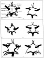 | 1,000 × 1,313 (155 KB) | Z8600021 (talk | contribs) | |
| 10:21, 16 November 2015 | 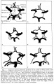 | 1,339 × 2,095 (407 KB) | Z8600021 (talk | contribs) | {{Historic Disclaimer}} Historic Embryology Papers ===Reference=== Gillilan, LA. [[Paper - Significant superficial anastomoses in the arterial blood supply to the human brain|Significant superficial anastomoses in the arterial blood supply to the... |
You cannot overwrite this file.


