File:Human Embryo 17.8mmCNS GIT.jpg
Original file (500 × 678 pixels, file size: 105 KB, MIME type: image/jpeg)
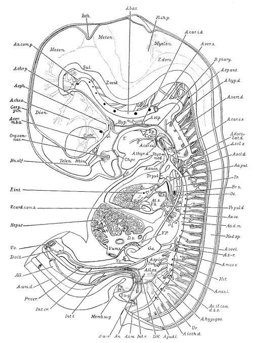
Human Embryo (17.8mm) Gastrointestinal Tract
Brain and Digestive System
Reconstruction to illustrate chiefly the interior of the brain and the spinal cord; the digestive system and its appendages; thc arterial system; the left atrium and ventricle of the heart, and in part the urogenital system of a 17.8 mm. hurrian embryo (H.E.C.839).
Original file name: Thyng100.jpg
The embryo external appearance and dimensions suggest that it is a Carnegie stage 19 embryo (Week 7, 48 - 51 days, 16 - 18 mm).
Reference: Thyng F.W., (1914) The anatomy of a 17.8mm human embryo. American Journal of Anatomy 14, 31-113
A reproduction of figure 104, page 153 of “Laboratory Textbook of Embryology,” Charles Sedgwick Minot, edition of 1910, published by P. Blakiston’s Son and Company, Philadelphia.
The American Journal of Anatomy, Vol.17, No.1 These drawings are based on studies of the Harvard Embryological Collection while he was in Minot's Lab in 1907-08.
He was also an anatomist at Northwestern University Medical School.
UNSW Embryology Link: Development History- 1914 - 17.8mm Human Embryo
- Dr Mark Hill 2009, UNSW Embryology ISBN: 978 0 7334 2609 4 - UNSW CRICOS Provider Code No. 00098G
File history
Yi efo/eka'e gwa ebo wo le nyangagi wuncin ye kamina wunga tinya nan
| Gwalagizhi | Nyangagi | Dimensions | User | Comment | |
|---|---|---|---|---|---|
| current | 14:36, 4 August 2009 | | 500 × 678 (105 KB) | MarkHill (talk | contribs) | Human Embryo (17.8mm) Gastrointestinal Tract Brain and Digestive System Reconstruction to illustrate chiefly the interior of the brain and the spinal cord; the digestive system and its appendages; thc arterial system; the left atrium and ventricle of th |
You cannot overwrite this file.
File usage
The following 4 pages use this file:


