File:Adult female fibroblast nuclear DNA 01.jpg
Original file (2,186 × 1,000 pixels, file size: 219 KB, MIME type: image/jpeg)
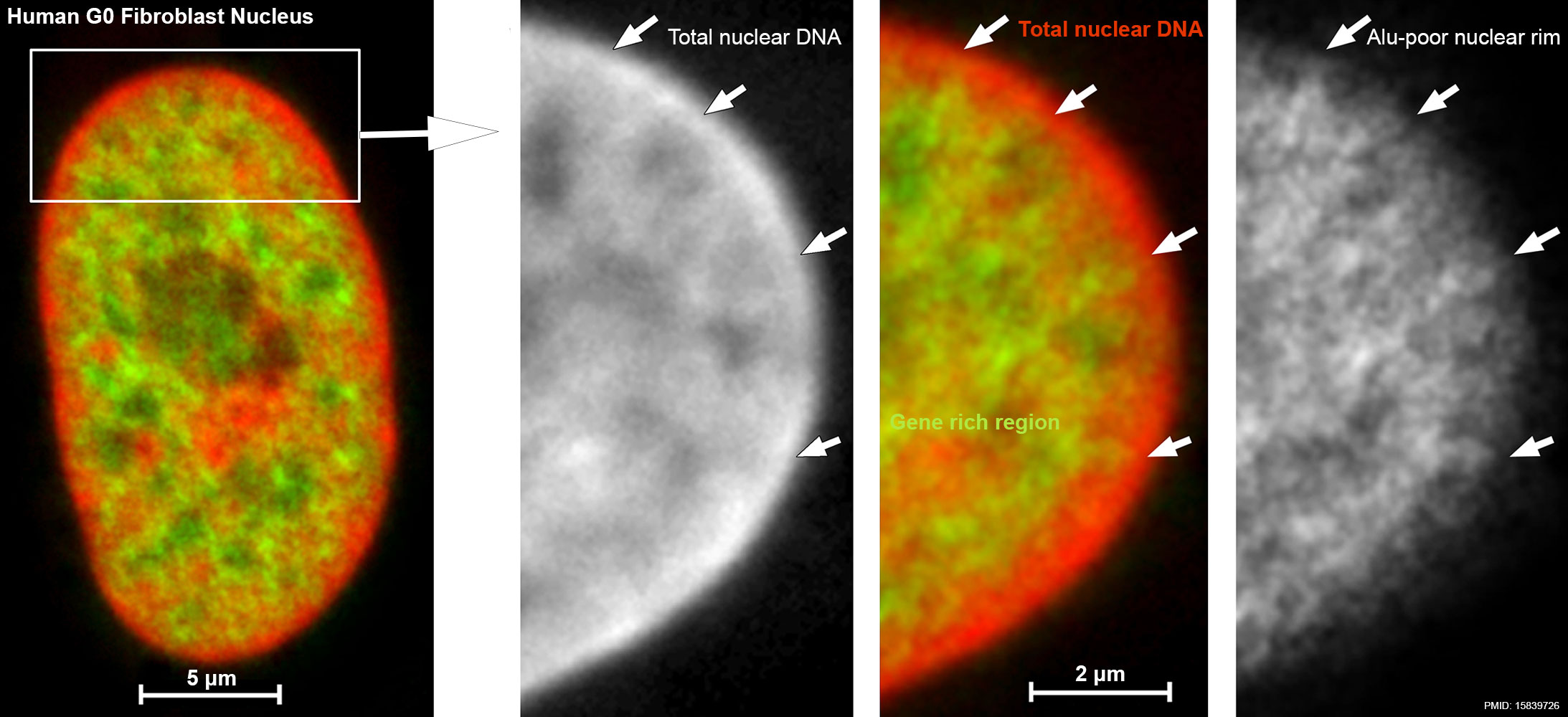
Adult female fibroblast nuclear DNA
Enlarged confocal mid-section through the human G0 fibroblast nucleus. Scale bar, 5 μm.
Righthand panels are enlargement of the boxed sector.
The color image in the middle reflects the merged images left (TOPRO-3 counterstaining, red) and right (Alu staining, green). Arrows indicate chromatin rich in Alu sequences expanding into the TOPRO-3-stained, Alu-poor nuclear rim. Scale bar, 2 μm.
- Genetics Links: Image - Female karyotype and nucleus gene locations | Image - Female karyotype | |Image - Female fibroblast nucleus gene locations | Cartoon - nucleus structure | Genetics
Reference
<pubmed>15839726</pubmed>| PLoS Biol.
Copyright
© 2005 Bolzer et al. This is an open-access article distributed under the terms of the Creative Commons Attribution License, which permits unrestricted use, distribution, and reproduction in any medium, provided the original work is properly cited.
Figure 7. doi:10.1371/journal.pbio.0030157.g007 PMID--15839726journal.pbio.0030157.g007.jpg panel D and E cropped, resized and relabelled from original figure.
File history
Yi efo/eka'e gwa ebo wo le nyangagi wuncin ye kamina wunga tinya nan
| Gwalagizhi | Nyangagi | Dimensions | User | Comment | |
|---|---|---|---|---|---|
| current | 13:58, 16 October 2014 | | 2,186 × 1,000 (219 KB) | Z8600021 (talk | contribs) |
You cannot overwrite this file.
File usage
The following page uses this file:


