File:Placental villi.jpg
From Embryology

Size of this preview: 750 × 600 pixels. Other resolution: 1,280 × 1,024 pixels.
Original file (1,280 × 1,024 pixels, file size: 199 KB, MIME type: image/jpeg)
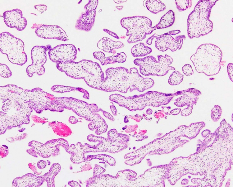
Human placental villi cross-section
Histology placenta, first trimester, human H&E
reproductive system, female, chorionic villi
original file name Ple20he.jpg
Image Source: UWA Blue Histology http://www.lab.anhb.uwa.edu.au/mb140/CorePages/FemaleRepro/femalerepro.htm
File history
Yi efo/eka'e gwa ebo wo le nyangagi wuncin ye kamina wunga tinya nan
| Gwalagizhi | Nyangagi | Dimensions | User | Comment | |
|---|---|---|---|---|---|
| current | 02:33, 1 April 2012 |  | 1,280 × 1,024 (199 KB) | Z8600021 (talk | contribs) | ==Human Placental Villi== * Villi are cover in shell of cytotrophoblast cells. * Villi core contains mainly mesenchyme cells. {{Placental villi histology}} {{Blue Histology}} original file name Ple20he.jpg Category:Human Embryo [[Category:Pla |
| 16:31, 3 August 2009 |  | 1,280 × 1,024 (250 KB) | MarkHill (talk | contribs) | Human placental villi cross-section Histology placenta, first trimester, human H&E reproductive system, female, chorionic villi original file name Ple20he.jpg Image Source: UWA Blue Histology http://www.lab.anhb.uwa.edu.au/mb140/CorePages/FemaleRepro/ |
You cannot overwrite this file.