File:Spermatozoa mitochondria morula.jpg

Original file (906 × 306 pixels, file size: 38 KB, MIME type: image/jpeg)
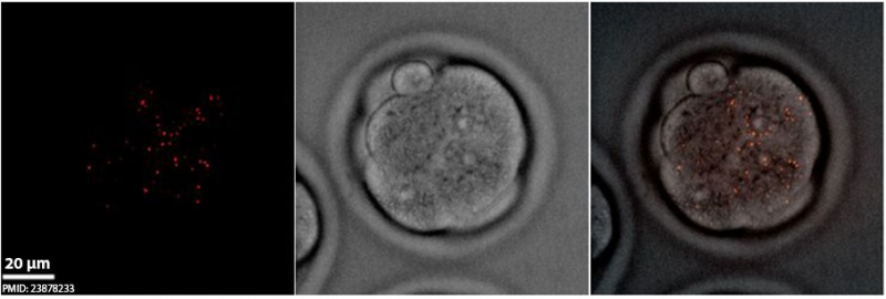
Spermatozoa Mitochondria in Early Mouse Embryos
Live cell fluorescence imaging of sperm mitochondria (red) in early embryos. Sperm mitochondria disaggregated and became restricted to only one blastomere during one-cell to four-cell stages. Sperm mitochondria distributed in several cells after the eight-cell stage and could be detected until morula stages.
Scale bars: 20 µm.
- Links: Mitochondria | Spermatozoa Development | Fertilization
Reference
<pubmed>23878233</pubmed>| Proc Natl Acad Sci U S A.
Copyright
Proceedings National Academy of Sciences (PNAS) Liberalization of PNAS copyright policy: Noncommercial use freely allowed Note original Author should be contacted for permission to reuse for Educational purposes. See also PNAS Author Rights and Permission FAQs
- Cozzarelli NR, Fulton KR, Sullenberger DM. Liberalization of PNAS copyright policy: noncommercial use freely allowed. Proc Natl Acad Sci U S A. 2004 Aug 24;101(34):12399. PMID15314225 "Our guiding principle is that, while PNAS retains copyright, anyone can make noncommercial use of work in PNAS without asking our permission, provided that the original source is cited."
Fig. 2. http://www.pnas.org/content/110/32/13038/F2.expansion.html Original image resized and relabeled.
File history
Yi efo/eka'e gwa ebo wo le nyangagi wuncin ye kamina wunga tinya nan
| Gwalagizhi | Nyangagi | Dimensions | User | Comment | |
|---|---|---|---|---|---|
| current | 14:35, 8 April 2014 | 906 × 306 (38 KB) | Z8600021 (talk | contribs) | ==Spermatozoa Mitochondria in Early Mouse Embryos== Live cell fluorescence imaging of sperm mitochondria (red) in early embryos. Sperm mitochondria disaggregated and became restricted to only one blastomere during one-cell to four-cell stages. Sperm ... |
You cannot overwrite this file.
File usage
The following 2 pages use this file: