File:Gray0784.jpg
Original file (851 × 600 pixels, file size: 137 KB, MIME type: image/jpeg)
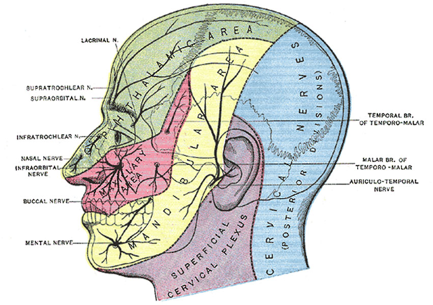
Fig. 784. Sensory Areas of the Head
Showing the general distribution of the three divisions of the fifth nerve (N. Trigeminus, Trifacial Nerve, CN V).
The trigeminal nerve is the largest cranial nerve and is the great sensory nerve of the head and face, and the motor nerve of the muscles of mastication. It emerges from the side of the pons, near its upper border, by a small motor and a large sensory root—the former being situated in front of and medial to the latter.
(Image Modified from Testut.)
(text modified from Gray's Anatomy)
- Gray's Images: Development | Lymphatic | Neural | Vision | Hearing | Somatosensory | Integumentary | Respiratory | Gastrointestinal | Urogenital | Endocrine | Surface Anatomy | iBook | Historic Disclaimer
| Historic Disclaimer - information about historic embryology pages |
|---|
 |
| iBook - Gray's Embryology | |
|---|---|

|
|
Reference
Gray H. Anatomy of the human body. (1918) Philadelphia: Lea & Febiger.
Cite this page: Hill, M.A. (2024, June 25) Embryology Gray0784.jpg. Retrieved from https://embryology.med.unsw.edu.au/embryology/index.php/File:Gray0784.jpg
- © Dr Mark Hill 2024, UNSW Embryology ISBN: 978 0 7334 2609 4 - UNSW CRICOS Provider Code No. 00098G
File history
Yi efo/eka'e gwa ebo wo le nyangagi wuncin ye kamina wunga tinya nan
| Gwalagizhi | Nyangagi | Dimensions | User | Comment | |
|---|---|---|---|---|---|
| current | 13:13, 15 May 2013 | | 851 × 600 (137 KB) | Z8600021 (talk | contribs) | ==Fig. 784. Sensory Areas of the Head== Showing the general distribution of the three divisions of the fifth nerve (N. Trigeminus, Trifacial Nerve, CN V). The trigeminal nerve is the largest cranial nerve and is the great sensory nerve of the head a... |
You cannot overwrite this file.
File usage
The following 3 pages use this file: