File:Anencephaly ultrasound.jpg
From Embryology
Size of this preview: 800 × 585 pixels. Other resolution: 900 × 658 pixels.
Original file (900 × 658 pixels, file size: 108 KB, MIME type: image/jpeg)
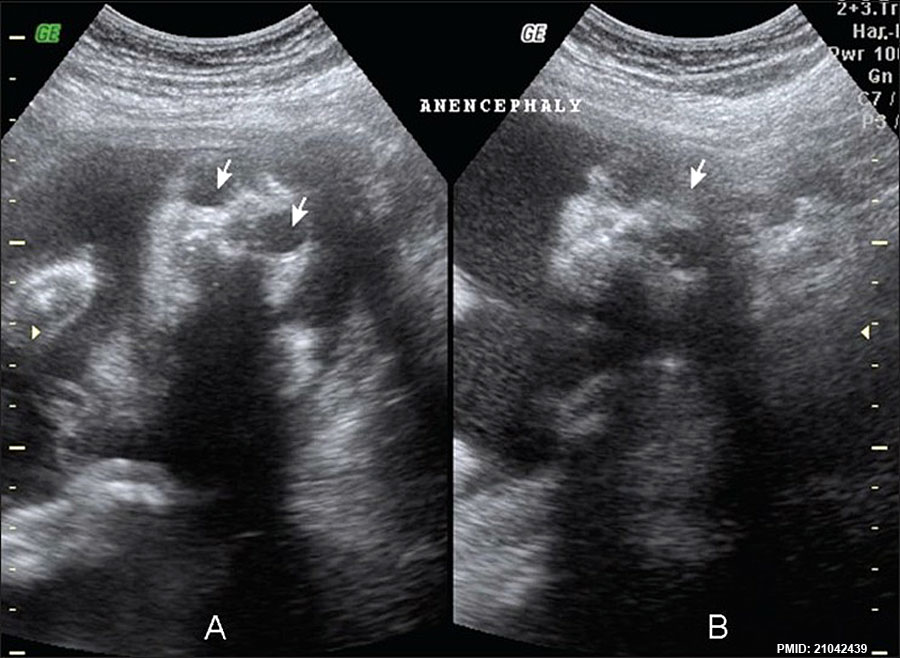
Anencephaly ultrasound
Prenatal ultrasound done at 18 weeks shows coronal images of the face and orbits with symmetric and complete absence of the cranial vault and brain.
- arrows in A - large and prominent orbits.
- arrows in B - complete absence of the cranial vault and brain.
Reference
Copyright
Figure 1 IndianJRadiolImaging_2010_20_3_174_69349_f4.jpg
File history
Yi efo/eka'e gwa ebo wo le nyangagi wuncin ye kamina wunga tinya nan
| Gwalagizhi | Nyangagi | Dimensions | User | Comment | |
|---|---|---|---|---|---|
| current | 14:10, 22 March 2013 | | 900 × 658 (108 KB) | Z8600021 (talk | contribs) | ==Anencephaly ultrasound== Prenatal ultrasound done at 18 weeks shows coronal images of the face and orbits with symmetric and complete absence of the cranial vault and brain. * arrows in A - large and prominent orbits. * arrows in B - complete absen... |
You cannot overwrite this file.


