File:Spleen histology 03.jpg
From Embryology
No higher resolution available.
Spleen_histology_03.jpg (450 × 600 pixels, file size: 83 KB, MIME type: image/jpeg)
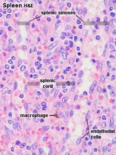
Spleen Histology - Cords and Sinuses
- Blood cells which are emptied into the splenic cords re-enter the blood vessels through the endothelium of the sinusoids.
- The endothelial cells are elongated (in cross section they may appear cuboidal) and oriented along the long axis of the sinusoids.
- The endothelium of the sinusoids has no junctional complexes and its basement membrane is incomplete.
- forming narrow circular bands around the endothelial cells with large intervening fenestrations.
- Macrophages ingest aged erythrocytes, platelets and other particulate matter as they pass through the splenic cords.
- The sinusoids continue into the veins of the pulp, which empty into thin-walled trabecular veins, which eventually coalesce to form the splenic vein.
Links: Histology | Histology Stains | Blue Histology images copyright Lutz Slomianka 1998-2009. The literary and artistic works on the original Blue Histology website may be reproduced, adapted, published and distributed for non-commercial purposes. See also the page Histology Stains.
Cite this page: Hill, M.A. (2026, October 5) Embryology Spleen histology 03.jpg. Retrieved from https://embryology.med.unsw.edu.au/embryology/index.php/File:Spleen_histology_03.jpg
- © Dr Mark Hill 2026, UNSW Embryology ISBN: 978 0 7334 2609 4 - UNSW CRICOS Provider Code No. 00098G
Original File name: Spl42he.jpg
File history
Yi efo/eka'e gwa ebo wo le nyangagi wuncin ye kamina wunga tinya nan
| Gwalagizhi | Nyangagi | Dimensions | User | Comment | |
|---|---|---|---|---|---|
| current | 20:30, 22 February 2012 | | 450 × 600 (83 KB) | Z8600021 (talk | contribs) | |
| 15:55, 21 February 2011 |  | 300 × 400 (51 KB) | S8600021 (talk | contribs) | ==Spleen Histology== Original File name: Spl42he.jpg {{Blue Histology}} Category:Spleen Category:Endocrine Category:Histology Category:Immune |
You cannot overwrite this file.


