File:Ultrasound12wk 3D image2.jpg
From Embryology
No higher resolution available.
Ultrasound12wk_3D_image2.jpg (362 × 264 pixels, file size: 8 KB, MIME type: image/jpeg)
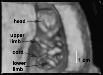
Ultrasound Fetus (12 week)
Ultrasound12wk 3D image2.jpg
Image from near end of movie showing ventral view of fetus head to top, upper limbs, lower limbs and umbilical cord visible.
Note the small cranial appearance, the cranial vault relatively under developed at this stage.
Compare this with the later 19 week ultrasound images.
Week 12 GA?
- Links: Ultrasound
File history
Yi efo/eka'e gwa ebo wo le nyangagi wuncin ye kamina wunga tinya nan
| Gwalagizhi | Nyangagi | Dimensions | User | Comment | |
|---|---|---|---|---|---|
| current | 20:57, 4 April 2010 | | 362 × 264 (8 KB) | S8600021 (talk | contribs) | Ultrasound12wk 3D image2.jpg |
You cannot overwrite this file.
File usage
The following 20 pages use this file:
- 2010 Foundations Lecture - Introduction to Human Development
- 2011 Lab 4
- ANAT2341 Lab 11 - Second Trimester
- ANAT2341 Lab 4 - Abnormal Placenta
- BGDA Practical 12 - Second Trimester
- BGDA Practical Placenta - Diagnostic Techniques
- Foundations Lecture - Introduction to Human Development
- Foundations Practical - Week 9 to 36
- Movies
- Pre-Medicine Program - Embryology
- Quicktime Ultrasound - Fetus 12 week
- Second Trimester
- Ultrasound
- Ultrasound - Fetus 12 week
- Talk:Flash Movies
- Template:Ultrasound 12wk movie
- Template:Ultrasound Movies
- Template:Ultrasound Movies - normal
- Template talk:Ultrasound Movies
- Help:Movies