File:Ovary-human-follicle.jpg
Original file (492 × 1,000 pixels, file size: 103 KB, MIME type: image/jpeg)
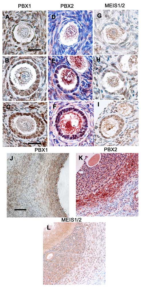
Immunohistochemical staining for PBX1, PBX2 and MEIS1/2 in human ovaries. (A, B, C, J) PBX1, (D, E, F, K) PBX2 and (G, H, I, L) MEIS1/2.
(A, D, G): primordial follicles.
(B, E, H): primary follicles.
(C, F, I): secondary follicles.
(J, K, L): preovulatory follicles. Figure 2E contains both primordial and primary follicles, and Figure 2I contains both primordial and secondary follicles
(scale bars: A,B,D,E,G,H = 9 um, C,F,I = 40 um, J,K,L = 350 um).
Reference
http://www.rbej.com/content/6/1/49
© 2008 Ota et al; licensee BioMed Central Ltd. This is an Open Access article distributed under the terms of the Creative Commons Attribution License (http://creativecommons.org/licenses/by/2.0), which permits unrestricted use, distribution, and reproduction in any medium, provided the original work is properly cited.
File history
Yi efo/eka'e gwa ebo wo le nyangagi wuncin ye kamina wunga tinya nan
| Gwalagizhi | Nyangagi | Dimensions | User | Comment | |
|---|---|---|---|---|---|
| current | 22:21, 24 August 2009 | | 492 × 1,000 (103 KB) | S8600021 (talk | contribs) | Immunohistochemical staining for PBX1, PBX2 and MEIS1/2 in human ovaries. (A, B, C, J) PBX1, (D, E, F, K) PBX2 and (G, H, I, L) MEIS1/2. (A, D, G): primordial follicles. (B, E, H): primary follicles. (C, F, I): secondary follicles. (J, K, L): preo |
You cannot overwrite this file.
File usage
There are no pages that use this file.