Category:Spinal Cord
From Embryology
This page lists UNSW Embryology content related to development of the spinal cord.
Pages in category 'Spinal Cord'
The following 36 pages are in this category, out of 36 total.
M
P
- Paper - Cell columns in the spinal cord of a human foetus of fourteen weeks (1941)
- Paper - Development of the innervation pattern in the upper limb of staged human embryos (1990)
- Paper - Factors Involved In The Formation Of The Filum Terminale
- Paper - The development and significance of the cell columns in the ventral horn of the cervical and upper thoracic spinal cord of the rabbit (1941)
- Paper - The early development of the meninges of the spinal cord in human embryos (1951)
- Paper - The structure of the spinal cord of the ostrich
R
- Template:Ref-Bardeen1903
- Template:Ref-DartShellshear1922
- Template:Ref-Hogg1945
- Template:Ref-Hoskins1914
- Template:Ref-Kunitomo1920
- Template:Ref-Miller1913
- Template:Ref-O’RahillyMuller1986
- Template:Ref-Romanes1941
- Template:Ref-Romanes1941a
- Template:Ref-Romanes1941b
- Template:Ref-ScharpenbergWindle1938
- Template:Ref-Sensenig1951
Media in category 'Spinal Cord'
The following 65 files are in this category, out of 65 total.
 Bailey385.jpg 787 × 683; 165 KB
 Bailey403.jpg 495 × 617; 118 KB
 Bailey407.jpg 793 × 695; 117 KB
 Caudal duplication syndrome.jpg 700 × 599; 47 KB
 Cervical vertebra.jpg 767 × 514; 71 KB
 Gray0664.jpg 696 × 500; 123 KB
 Gray0666.jpg 237 × 1,000; 46 KB
 Gray0666new.jpg 600 × 551; 47 KB
 Gray0670.jpg 800 × 504; 67 KB
 Gray0671.jpg 347 × 900; 54 KB
 Gray0675.jpg 615 × 600; 69 KB
 Gray0770.jpg 700 × 275; 68 KB
 Gray0804.jpg 550 × 700; 75 KB
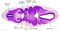
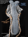
 Human Stage22 spinal cord01.jpg 1,044 × 889; 265 KB
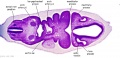
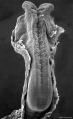
 Human Stage22 spinal cord02.jpg 1,044 × 889; 290 KB
 Human week 10 fetus 11.jpg 1,200 × 900; 304 KB
 Keith1921 fig064.jpg 908 × 565; 96 KB
 Keith1921 fig065.jpg 1,200 × 890; 219 KB
 Keith1921 fig066.jpg 1,000 × 1,067; 100 KB
 Keith1921 fig067.jpg 1,200 × 722; 150 KB
 Keith1921 fig068.jpg 903 × 784; 170 KB
 Keith1921 fig073a.jpg 884 × 822; 162 KB
 Keith1921 fig074.jpg 1,045 × 602; 134 KB
 Keith1921 fig075.jpg 856 × 636; 125 KB
 Keith1921 fig076.jpg 1,207 × 729; 265 KB
 Keith1921 fig077.jpg 629 × 640; 72 KB
 Keith1921 fig079.jpg 741 × 839; 109 KB
 Kollmann646.jpg 1,000 × 606; 122 KB
 Kollmann647.jpg 897 × 926; 188 KB
 McMurrich1930 fig81.jpg 1,280 × 796; 137 KB
 Mouse- spinal cord axons.jpg 600 × 693; 127 KB
 Neural tube SHH patterning cartoon.jpg 458 × 594; 92 KB
 Rugh 119.jpg 842 × 800; 127 KB
 Rugh 132.jpg 735 × 1,000; 195 KB
 Rugh 133.jpg 1,000 × 707; 141 KB
 Sensenig1951 plate01.jpg 1,979 × 2,591; 1.35 MB
 Sensenig1951 plate02.jpg 2,078 × 2,619; 1.52 MB
 Sensenig1951 plate03.jpg 1,963 × 2,664; 1.33 MB
 Sensenig1951 plate04.jpg 1,990 × 2,627; 1.29 MB
 Spinal cord delta notch model.png 599 × 457; 171 KB
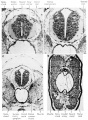
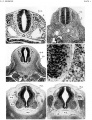
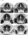
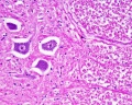
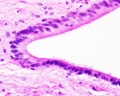
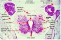
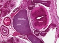
 Spinal cord histology 01.jpg 480 × 600; 116 KB
 Spinal cord histology 02.jpg 480 × 600; 121 KB
 Spinal cord histology 03.jpg 480 × 600; 103 KB
 Spinal cord histology 04.jpg 480 × 600; 119 KB
 Spinal cord histology 05.jpg 1,280 × 1,024; 463 KB
 Spinal cord histology 06.jpg 1,280 × 1,024; 318 KB
 Spinal cord histology 07.jpg 1,280 × 1,024; 365 KB
 Spinal cord histology 08.jpg 1,280 × 1,024; 418 KB
 Spinal cord histology 09.jpg 1,280 × 1,024; 227 KB
 Spinal cord histology 10.gif 480 × 600; 456 KB
 Spinal cord histology 11.jpg 481 × 600; 117 KB
 Spinal cord histology 12.jpg 480 × 600; 130 KB
 Spinal cord tracts.png 1,000 × 430; 204 KB
 Stage 13 image 007.jpg 1,000 × 514; 93 KB
 Stage 13 image 022.jpg 1,000 × 473; 101 KB
 Stage 13 image 023.jpg 1,000 × 544; 110 KB
 Stage 13 image 057.jpg 1,000 × 511; 99 KB
 Stage 13 image 058.jpg 1,000 × 481; 94 KB
 Stage 13 image 059.jpg 1,000 × 513; 92 KB
 Stage 13 image 060.jpg 1,000 × 486; 96 KB
 Stage 13 image 061.jpg 1,000 × 600; 101 KB
 Stage 22 image 176.jpg 1,000 × 659; 223 KB
 Stage10 sem6 annotated.jpg 720 × 960; 122 KB
 Stage10 sem6.jpg 614 × 1,000; 57 KB
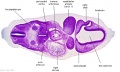
 Stage22 vertebra and spinal cord 1.jpg 1,000 × 725; 344 KB


