File:Stage8 sem6.jpg
Original file (1,000 × 698 pixels, file size: 87 KB, MIME type: image/jpeg)
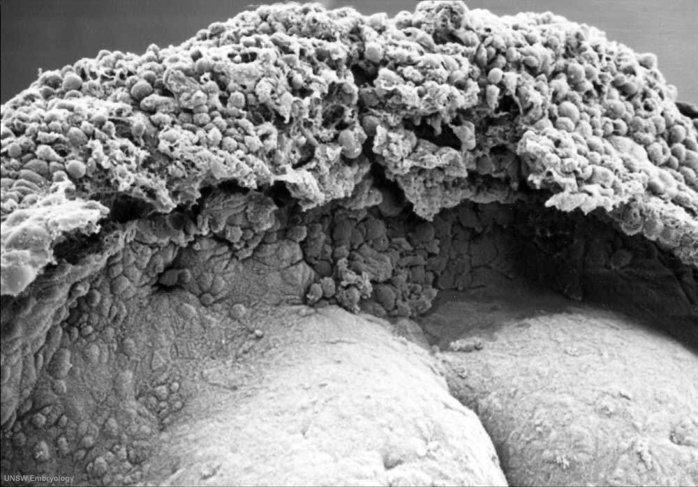
Human embryo (Stage 8, day 18)
Scanning EM dorsal view (cut amnion) showing detailed view of the prechordal region.
Selected region from the image selected rostral (cranial) region, which is in turn the rostral region of the entire embryonic disc image.
Note the rostro-caudal axis (top to bottom), the growing brain region of the neural plate (top) and shape of the folding embryonic disc.
Primitive node and streak still visible below the region where the posterior (caudal) neuropore will be located.
Note the cut edge of amniotic sac with two layers (inner ectoderm, outer extra-embryonic mesoderm)
Original image name: PresomiteStage8day18Dorsal04.jpg
Image Source: Scanning electron micrographs of the Carnegie stages of the early human embryos are reproduced with the permission of Prof Kathy Sulik, from embryos collected by Dr. Vekemans and Tania Attié-Bitach. Images are for educational purposes only and cannot be reproduced electronically or in writing without permission.
File history
Yi efo/eka'e gwa ebo wo le nyangagi wuncin ye kamina wunga tinya nan
| Gwalagizhi | Nyangagi | Dimensions | User | Comment | |
|---|---|---|---|---|---|
| current | 09:36, 22 August 2009 | | 1,000 × 698 (87 KB) | S8600021 (talk | contribs) | Human embryo (Stage 8, day 18) Scanning EM dorsal view (cut amnion) showing detailed view of the prechordal region. Note the rostro-caudal axis (top to bottom), the growing brain region of the neural plate (top) and shape of the folding embryonic disc. |
You cannot overwrite this file.
File usage
The following 4 pages use this file: