File:Human-oocyte to blastocyst.jpg
Human-oocyte_to_blastocyst.jpg (600 × 402 pixels, file size: 49 KB, MIME type: image/jpeg)
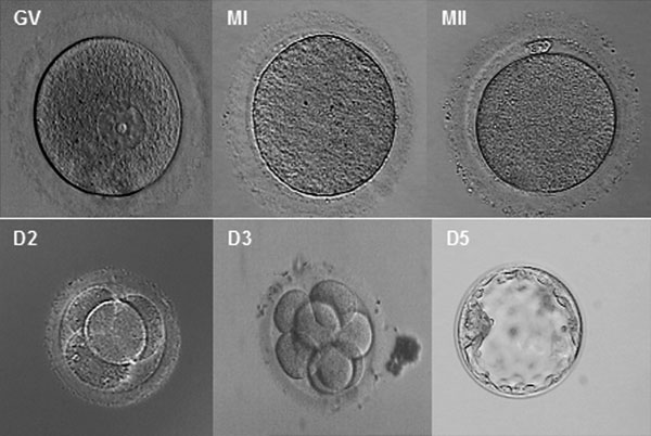
Morphology of human oocytes and embryos.
GV - germinal vesicle oocyte (GV)
MI - metaphase I oocyte
MII - metaphase II oocyte
D2 - 4-cell embryo
D3 - 8-cell embryo
D5 - blastocyst
- MII oocytes had been matured in vitro after donation at GV stage. D2, D3, D5 embryos were all matured in vitro.
Note - Original image has been modified to remove array data and colour. See also File:Human-oocyte.jpg
Pone.0007844.g004.jpg
http://www.ncbi.nlm.nih.gov/pmc/articles/PMC2773928/figure/pone-0007844-g004/
Related Images: Human oocyte to blastocyst | Germinal vesicle oocyte (GV) | Metaphase I oocyte | Metaphase II oocyte | Day 2 | Day 3 | Day 5
Citation: <pubmed>19924284</pubmed>| PMC2773928 | PLoS One
PLoS One. 2009; 4(11): e7844. Published online 2009 November 16. doi: 10.1371/journal.pone.0007844.
PMCID: PMC2773928
Copyright Zhang et al. This is an open-access article distributed under the terms of the Creative Commons Attribution License, which permits unrestricted use, distribution, and reproduction in any medium, provided the original author and source are credited.
File history
Yi efo/eka'e gwa ebo wo le nyangagi wuncin ye kamina wunga tinya nan
| Gwalagizhi | Nyangagi | Dimensions | User | Comment | |
|---|---|---|---|---|---|
| current | 13:09, 5 April 2010 | | 600 × 402 (49 KB) | S8600021 (talk | contribs) | Morphology of human oocytes and embryos. GV - germinal vesicle oocyte (GV) MI - metaphase I oocyte MII - metaphase II oocyte D2 - 4-cell embryo D3 - 8-cell embryo D5 - blastocyst MII oocytes had been matured in vitro after donation at GV stage. D2 |
You cannot overwrite this file.
File usage
The following 16 pages use this file:
- 2010 Foundations Lecture - Introduction to Human Development
- BGDA Practical 3 - Early Cell Division
- Carnegie stage 1
- Pre-Medicine Program - Embryology
- Week 1
- File:Human-oocyte.jpg
- File:Human-oocyte to blastocyst.jpg
- File:Human embryo day 2.jpg
- File:Human embryo day 3.jpg
- File:Human embryo day 5.jpg
- File:Human embryo day 5 label.gif
- File:Human embryo day 5 label.jpg
- File:Human embryo day 5 label2.jpg
- File:Human oocyte-metaphase I.jpg
- File:Human oocyte-metaphase II.jpg
- Template:Human oocyte to blastocyst


