File:Fetal pineal gland 01.jpg
Original file (700 × 603 pixels, file size: 67 KB, MIME type: image/jpeg)
Fetal Pineal Gland Anatomy
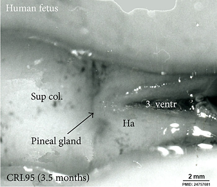
Superior (dorsal) view of the diencephalic-mesencephalic area of a 3.5-month-old human fetus.
The third ventricle (3 ventr) without pial covering is seen to the right in the micrograph. The small pineal gland is a small protuberance (arrow) and merging via the broad stalk with the habenula (Ha). Sup col.: superior colliculus. Bar = 2 mm.
- Links: Pineal Development | Fetal Development
Reference
<pubmed>24757681</pubmed>| PMC3976832 | Biomed Res Int.
Copyright
© 2014 Morten Møller et al. This is an open access article distributed under the Creative Commons Attribution License, which permits unrestricted use, distribution, and reproduction in any medium, provided the original work is properly cited.
Figure 3: 868567.fig.003.jpg http://www.hindawi.com/journals/bmri/2014/868567/fig3 Figure adjusted in size, sharpness and labelling.
File history
Yi efo/eka'e gwa ebo wo le nyangagi wuncin ye kamina wunga tinya nan
| Gwalagizhi | Nyangagi | Dimensions | User | Comment | |
|---|---|---|---|---|---|
| current | 10:05, 13 September 2014 | | 700 × 603 (67 KB) | Z8600021 (talk | contribs) | Superior (dorsal) view of the diencephalic-mesencephalic area of a 3.5-month-old human fetus. The third ventricle (3 ventr) without pial covering is seen to the right in the micrograph. The small pineal gland is a small protuberance (arrow) and mergi... |
You cannot overwrite this file.
File usage
The following 4 pages use this file:


