File:Spaulding-plate04.jpg
Original file (767 × 1,048 pixels, file size: 98 KB, MIME type: image/jpeg)
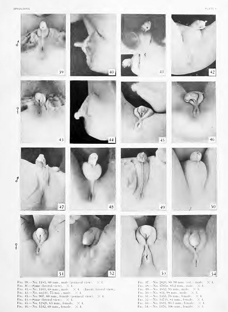
Plate 4
Fig. 39. No. 1183, 60 mm., male (perineal v-iew). X 4.
Fig. 40. No. 1183 Same (lateral view) . X 4.
Fig. 41. No. 1163, 68 mm., male. X 4. (Insert, lateral
Fig. 42. Xo. nn34fc, 75 mm, male. X 4.
Fig. 43. No. 907, 60 mm., female (perineal view). X 4.
Fig. 44. No. 907, Same (lateral view). X 4.
Fig. 45. No. 1282b, 65 mm., female. X 4.
Fig. 46. No. 1542, 69 mm., female. X 4.
Fig. 47. No. 2026, 80-90 mm. (est.), male.
Fig. 48. No. 1705a, 83.2 mm., male. X 4.
Fig. 49. No. 1852, 95 mm., male. X 3.
Fig. 50. No. 834, 98 mm., male. X 3.
Fig. 51. No. 1455, 78 mm., female. X 4.
Fig. 52. No. 1474b, 84 mm., female. X 4.
Fig. 53. No. 1831, 93.5 mm., female. X 4.
Fig. 54. No. 1476, 100 mm., female. X 4.
- Figure Links: Text | Text Figure 1 | Text Figure 2 | Plate 1 | Fig. 1 | Fig. 2 | Fig. 3 | Fig. 4 | Fig. 5 | Fig. 6 | Plate 2 | Fig. 7 | Fig. 8 | Fig. 9 | Fig. 10 | Fig. 11 | Fig. 12 | Fig. 13 | Fig. 14 | Fig. 15 | Fig. 16 | Fig. 17 | Fig. 18 | Fig. 19 | Fig. 20 | Fig. 21 | Fig. 22 | Plate 3 | Fig. 23 | Fig. 24 | Fig. 25 | Fig. 26 | Fig. 27 | Fig. 28 | Fig. 29 | Plate 4 | Fig. 30 | Fig. 31 | Fig. 32 |Fig. 33 | Fig. 34 | Fig. 35 | Fig. 36 | Fig. 37 | Fig. 38 | Fig. 39 | Fig. 40 | Fig. 41 | Fig. 42 | Fig. 43 | Fig. 44 | Fig. 45 | Fig. 46 | Fig. 47 | Fig. 48 | Fig. 49 | Fig. 50 | Fig. 51 | Fig. 52 | Fig. 53 | Fig. 54
| Historic Disclaimer - information about historic embryology pages |
|---|
 |
Reference
Spaulding MH. The development of the external genitalia in the human embryo. (1921) Contrib. Embryol., Carnegie Inst. Wash. Publ. 81, 13: 69 – 88.
Cite this page: Hill, M.A. (2024, June 20) Embryology Spaulding-plate04.jpg. Retrieved from https://embryology.med.unsw.edu.au/embryology/index.php/File:Spaulding-plate04.jpg
- © Dr Mark Hill 2024, UNSW Embryology ISBN: 978 0 7334 2609 4 - UNSW CRICOS Provider Code No. 00098G
| Historic Disclaimer - information about historic embryology pages |
|---|
|
File history
Yi efo/eka'e gwa ebo wo le nyangagi wuncin ye kamina wunga tinya nan
| Gwalagizhi | Nyangagi | Dimensions | User | Comment | |
|---|---|---|---|---|---|
| current | 16:48, 26 March 2011 | | 767 × 1,048 (98 KB) | S8600021 (talk | contribs) | ==Plate 4== {{Template:Historic Disclaimer}} ==Reference== The development of the external genitalia in the human embryo By Milo Herrick Spaulding, Of the University of Montmxa, Stale College of Agriculture, Bozeman. With four plates and two text-fi |
You cannot overwrite this file.
File usage
The following 2 pages use this file: