File:Licata1954 fig16.jpg

Original file (1,000 × 1,212 pixels, file size: 297 KB, MIME type: image/jpeg)
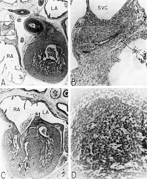
Fig. 16 Photomierographs showing parts of the developing sinoventricular conduction system in the heart of a 31.5 mm embryo
A. Section (X 27) passing through right atrium near level of entrance of superior vcna cava. Abbreviations: LA, left atrium; RA, right atrium.
B. Area. indicated by rectangle in A, photographed at higher magnification (X 125) to show the primordium of the sino-atrial node. Note the position of the nodal artery. Abbreviations: NA, nodal artery; SA, sino-atrial node; Sh, connective—tissue sheath; SVC, superior vena cava.
C. Section (X 20) through the interventricular septum from same heart as that shown in A. LA, left atrium; RA, right atrium.
D. Area indicated by rectangle in C (X 208) to show the main comluction bundle of His.
- Links: fig 1 | fig 2 | fig 3 | fig 4 | fig 5 | fig 6 | fig 7 | fig 8 | fig 9 | fig 10 | fig 11 | fig 12 | fig 13 | fig 14 | fig 15 | fig 16 | fig 16a | fig 16b | fig 16c | fig 16d | 1954 Licata | Historic Papers | Heart Development
Reference
Licata RH. The human embryonic heart in the ninth week. (1954) Amer. J Anat., 94: 73-125. PMID 13124266
Cite this page: Hill, M.A. (2024, June 21) Embryology Licata1954 fig16.jpg. Retrieved from https://embryology.med.unsw.edu.au/embryology/index.php/File:Licata1954_fig16.jpg
- © Dr Mark Hill 2024, UNSW Embryology ISBN: 978 0 7334 2609 4 - UNSW CRICOS Provider Code No. 00098G
File history
Yi efo/eka'e gwa ebo wo le nyangagi wuncin ye kamina wunga tinya nan
| Gwalagizhi | Nyangagi | Dimensions | User | Comment | |
|---|---|---|---|---|---|
| current | 12:01, 5 March 2017 | | 1,000 × 1,212 (297 KB) | Z8600021 (talk | contribs) | |
| 12:00, 5 March 2017 | 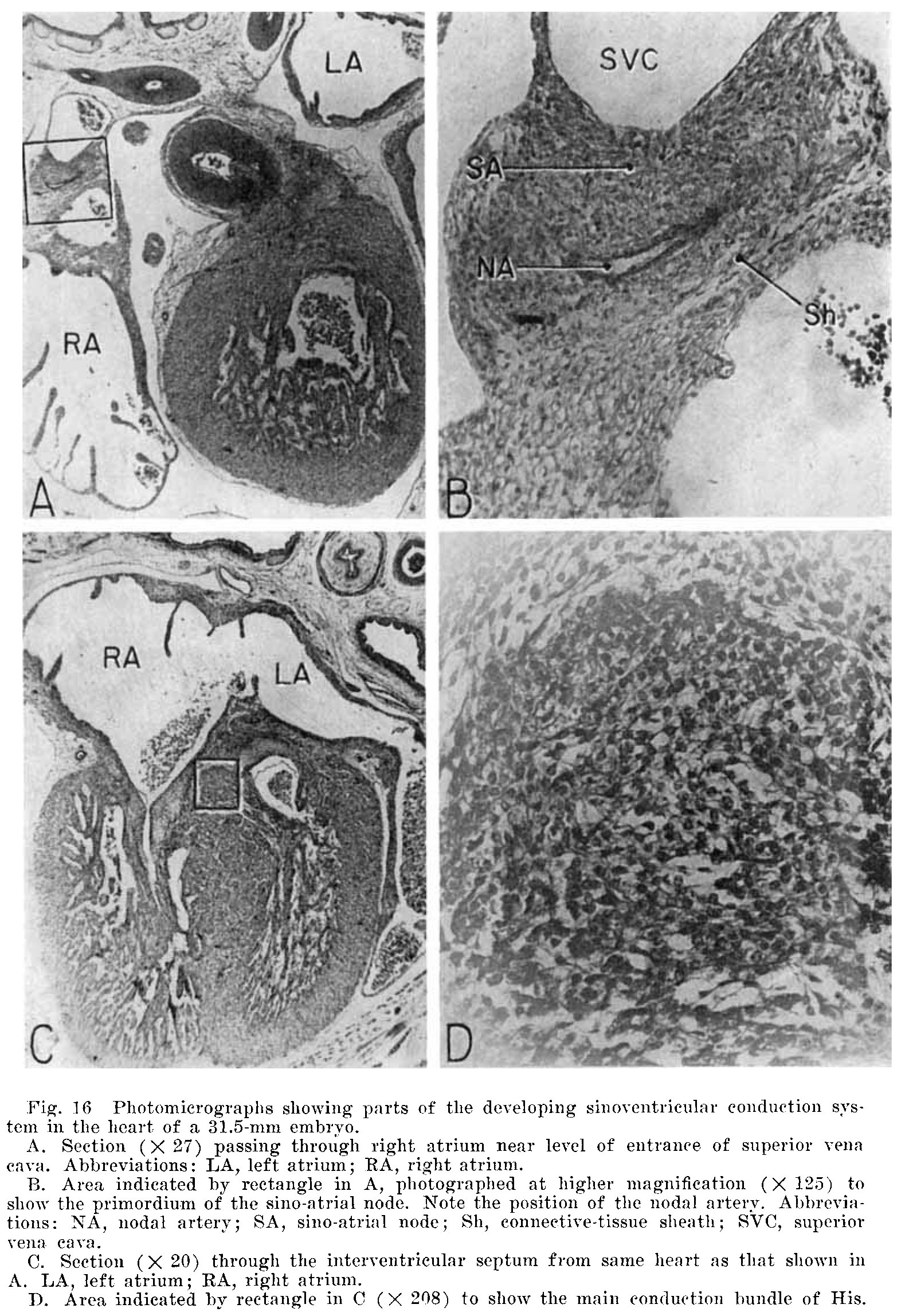 | 1,579 × 2,295 (746 KB) | Z8600021 (talk | contribs) |
You cannot overwrite this file.
File usage
The following 4 pages use this file: