Talk:Draft 2016

From Embryology
Revision as of 18:24, 26 February 2016 by Z8600021 (talk | contribs)

Lymph Nodes

Lymph Node Anatomy

Lymph Node Anatomy
  • Location throughout the entire body - concentrated in axilla, groin, lung, gastrointestinal tract mesenteries
  • Small (1 mm - 2 cm) encapsulated organ (diffuse lymphoid tissue - no capsule)
  • Antigen transformed lymphocytes from the blood
  • In lymph vessel pathways “filter” lymph
    • Afferent - towards node (A - arrives at the node)
    • Efferent - away from node (E - exits the node)

Gastrointestinal tract intestine immune cartoon 01.jpg

Mesenteric lymph nodes

Lymphatic-system-overview.jpg

Lymph Node Functions

Lymph Node Functions 
  • In lymph vessel pathways “filter” lymph
  • Immune - detect infections from peripheral tissues
    • skin, respiratory tract, gastrointestinal tract, etc.
  • secondary lymphoid organ
  • return extracellular fluid to circulation
Lymph node structure.jpg

Lymph Node Structure

Lymph Node Structure 
Lymph node cartoon.jpg
Lymph node cartoon 02.jpg

Lymph Node Histology

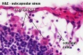
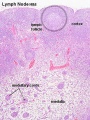
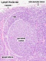
Lymph Node Histology 
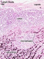
  • Capsule (thin) with trabecular or septa (dense connective tissue)

Lymph_node_cartoon_01.jpg


Fetal thymus Young medulla Young cortex
Fetal thymus.jpg Thymus - young 01.jpg Thymus - young 02.jpg


Lymph Node Cartoons: Detailed structure | Cartoon with Histology | Lymphocyte traffic | Simple structure | Simple node anatomy | Wiki node image | Internal structure | Mesenteric lymph node | Histology | Gallery | Lymph Node Development

Lymph Nodes

Lymph Node Structure

Lymph_node_cartoon_01.jpg

Connective Tissue

  • Capsule - dense connective tissue (irregular CT, some adipocytes))
  • Trabeculae - dense connective tissue
  • Reticular Tissue - Reticular cells and fibers, supporting meshwork (collagen type III)
    • Reticular cell produces reticular fibers (collagen type III) and surrounds the fibers with its cytoplasm
    • reticular fibbers can also be produced by fibroblasts
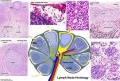

Lymphocyte (T and B) Traffic

  1. Enter from high endothelial venules (HEVs also called post-capillary venules)
  2. Spend 8 to 24 h in the lymph node interstitium.
  3. Enter a network of medullary sinuses.
  4. Drain from sinuses into efferent lymphatic vessels.


Important - Please note that the lymphocytes exit from the node by efferent lymphatic vessels.

See Figure

See also Image - Cell Trafficking into and out of Lymph Nodes.

Mouse adult lymph node 07.jpg
 ‎‎Lymph Node 7
Page | Play

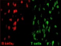
T and B motility

Mouse adult lymph node 08.jpg
 ‎‎Lymph Node 8
Page | Play

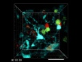
T and B interaction

Lymph node histology 05.jpg

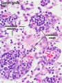
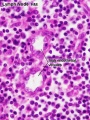
High Endothelial Venules

See also Lymphocyte Migration at High Endothelial Venule Model

Links: Immunobiology - Figure 1.8. Organization of a lymph node