File:Human embryo day 18.jpg
Original file (531 × 800 pixels, file size: 80 KB, MIME type: image/jpeg)
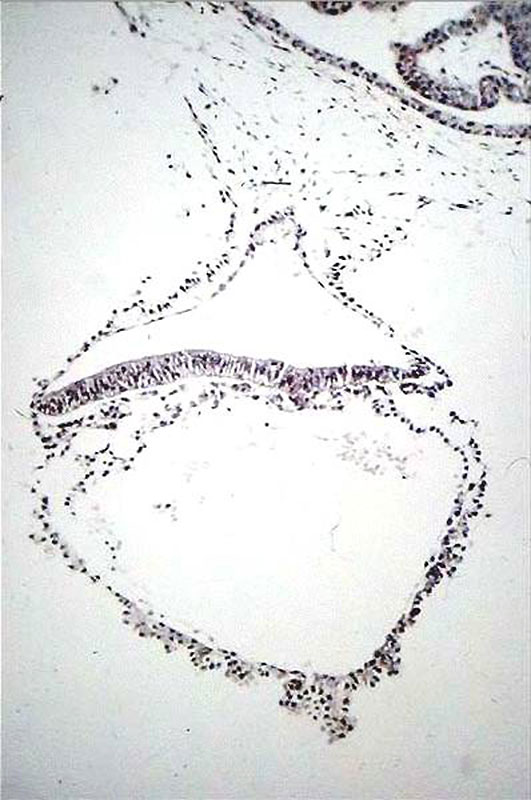
Human embryo day 18
Approximate cross-section of a different, 18 day human conceptus.
Identify the 3 cavities: yolk sac, amniotic sac and chorionic sac.
Identify the 3 layers of the trilaminar embryo: ectoderm (columnar cells), intraembryonic mesoderm (mesenchymal cells, endodermal cells (cuboidal single layer).
Identify the extraembryonic mesoderm which surrounds the yolk and amniotic sacs, and continues to form the connecting stalk, where it then continues to line the chorionic sac. Extra- and intra-embryonic mesoderm merge along the perimeter of the embryonic disc.
Identify primitive groove with dense cluster of primitive streak cells below it.
At the top right of the micrograph is a secondary chorionic villus, lying in an empty maternal intervillus space. The villous has an outer layer of syncytiotrophoblast (smaller, darker nuclei), an intermediate layer of cytotrophoblast and a core of mesoblast (extra-embryonic mesoderm).
(Image: Nishimura etal., 1977)
File history
Yi efo/eka'e gwa ebo wo le nyangagi wuncin ye kamina wunga tinya nan
| Gwalagizhi | Nyangagi | Dimensions | User | Comment | |
|---|---|---|---|---|---|
| current | 22:48, 8 October 2015 | | 531 × 800 (80 KB) | Z8600021 (talk | contribs) | |
| 21:38, 9 Mayıs 2010 |  | 398 × 600 (38 KB) | S8600021 (talk | contribs) | Human embryo day 18 Category:Week 3 |
You cannot overwrite this file.
File usage
The following 7 pages use this file:


