File:Papanicolaou1933-plate08.jpg
Original file (1,556 × 2,000 pixels, file size: 432 KB, MIME type: image/jpeg)
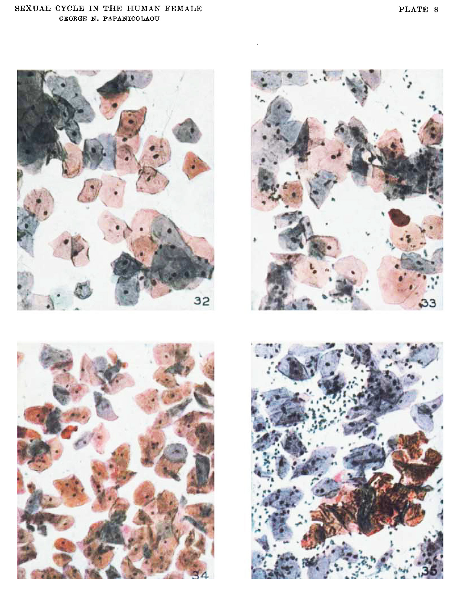
Plate 8 Photomicrographs of human vaginal smears colored by hand as in stained smears
Photomicrographs of human vaginal smears colored by hand as in stained smears, in order to show the relative number of cornified cells (stained red).
32 Twelfth day. Typical copulative smear with leucopenia. Same as figure 13.
33 Ovulative type of smear. Fourteenth day. Oc. 3,0bj. 8, B. 19.
34 Fifteenth day. Post-ovulative smear with continued high cornification and Ieueopenia. Same as figure 17.
35 Twenty-third day. Typical premenstrual smear. Same specimen as in figure 18. 0c. 6, Obj. 16, B. 23.
Reference
Papanicolaou GN. The Sexual Cycle in the Human Female as revealed by Vaginal Smears. Am J Anat. 1933;52: 519–637.
Cite this page: Hill, M.A. (2024, June 21) Embryology Papanicolaou1933-plate08.jpg. Retrieved from https://embryology.med.unsw.edu.au/embryology/index.php/File:Papanicolaou1933-plate08.jpg
- © Dr Mark Hill 2024, UNSW Embryology ISBN: 978 0 7334 2609 4 - UNSW CRICOS Provider Code No. 00098G
File history
Yi efo/eka'e gwa ebo wo le nyangagi wuncin ye kamina wunga tinya nan
| Gwalagizhi | Nyangagi | Dimensions | User | Comment | |
|---|---|---|---|---|---|
| current | 12:39, 8 September 2015 | | 1,556 × 2,000 (432 KB) | Z8600021 (talk | contribs) |
You cannot overwrite this file.
File usage
The following 2 pages use this file: