File:Measles virus.jpg
Measles_virus.jpg (700 × 535 pixels, file size: 91 KB, MIME type: image/jpeg)
Measles virus
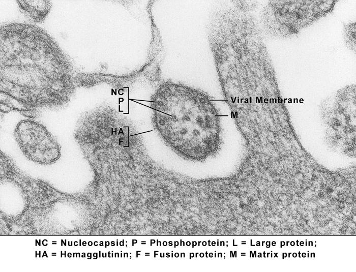
This transmission electron micrograph (TEM) revealed the ultrastructural appearance of a virus particle, or “virion”, of the measles virus. The measles virus is a paramyxovirus, of the genus Morbillivirus.
The viral particle is 100-200 nm in diameter, with a core of single-stranded RNA, and is closely related to the Rinderpest and canine distemper viruses. Two membrane envelope proteins are important in pathogenesis. They are the F (fusion) protein, which is responsible for fusion of virus and host cell membranes, viral penetration, and hemolysis, and the HA (hemagglutinin) protein, which is responsible for adsorption of virus to cells.
Copyright
None - This image is in the public domain and thus free of any copyright restrictions. As a matter of courtesy we request that the content provider be credited and notified in any public or private usage of this image.
Content Providers(s): CDC/ Brian W.J. Mahy Original Image name: 12733_lores.jpg http://phil.cdc.gov/PHIL_Images/12733/12733_lores.jpg]
File history
Click on a date/time to view the file as it appeared at that time.
| Date/Time | Thumbnail | Dimensions | User | Comment | |
|---|---|---|---|---|---|
| current | 11:35, 7 November 2011 |  | 700 × 535 (91 KB) | S8600021 (talk | contribs) | ==Measles virus== This transmission electron micrograph (TEM) revealed the ultrastructural appearance of a virus particle, or “virion”, of the measles virus. The measles virus is a paramyxovirus, of the genus Morbillivirus. The viral particle is 100 |
You cannot overwrite this file.
File usage
The following 2 pages use this file:
