File:Mouse-2 cell 01.jpg
Mouse-2_cell_01.jpg (400 × 398 pixels, file size: 14 KB, MIME type: image/jpeg)
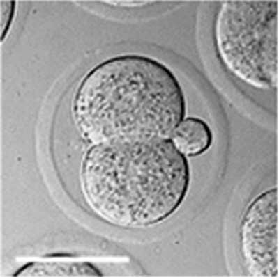
Mouse Embryo Blastomeres
Two blastomeres within the zone pellucida. Polar body visible to the right.
Scale bar 50 µm.
- Image Links: zygote | blastomeres | morula | early blastocyst | hatched blastocyst | Mouse Development | Fertilization | Morula | Blastocyst
Reference
<pubmed>20405021</pubmed>| PMC2854157 | PLoS
Editor: Mai Har Sham, The University of Hong Kong, China
Received: December 3, 2009; Accepted: March 23, 2010; Published: April 13, 2010
Copyright: © 2010 Valley et al. This is an open-access article distributed under the terms of the Creative Commons Attribution License, which permits unrestricted use, distribution, and reproduction in any medium, provided the original author and source are credited.
File history
Yi efo/eka'e gwa ebo wo le nyangagi wuncin ye kamina wunga tinya nan
| Gwalagizhi | Nyangagi | Dimensions | User | Comment | |
|---|---|---|---|---|---|
| current | 13:35, 17 April 2010 |  | 400 × 398 (14 KB) | S8600021 (talk | contribs) |
You cannot overwrite this file.
File usage
The following 2 pages use this file:
