Category:Electron Micrograph
From Embryology
This page lists UNSW Embryology content related to electron micrographs, see also the related category Scanning Electron Micrograph.
Pages in category 'Electron Micrograph'
The following 22 pages are in this category, out of 22 total.
H
P
- Paper - Cell-to-cell communication and ovulation - A study of the cumulus-oocyte complex
- Paper - Electron microscopy of the sperm tail - results obtained with a new fixative
- Paper - Fine structure of the human ovum in the pronuclear stage
- Paper - Studies on the fine structure of the mammalian testis 1
- Paper - Studies on the human oocyte and its follicle 1
- Template:Placenta EM links
R
Media in category 'Electron Micrograph'
The following 200 files are in this category, out of 201 total.
(previous page) (next page) 1918 influenza virus virions EM.jpg 700 × 743; 82 KB
 2009 influenza virus virions EM.jpg 700 × 676; 126 KB
 Adenovirus.jpg 700 × 499; 45 KB
 Amphibian oocyte transcription.jpg 1,200 × 841; 475 KB
 Apoptosis and necrosis.jpg 800 × 400; 60 KB
 Avian influenza virion.jpg 320 × 240; 12 KB
 B lymphocyte EM08.jpg 1,000 × 730; 111 KB
 B lymphocyte EM09.jpg 673 × 1,000; 89 KB
 B lymphocyte EM10.jpg 671 × 1,000; 92 KB
 B lymphocytes EM08-10.jpg 677 × 996; 124 KB
 Blood capillary EM 01.jpg 1,107 × 714; 260 KB
 Blood capillary EM 02.jpg 600 × 600; 99 KB
 Blood capillary EM 03.jpg 1,560 × 1,230; 441 KB
 Blood capillary EM 04.jpg 1,560 × 1,230; 462 KB
 Blood capillary EM 05.jpg 1,015 × 800; 205 KB
 Blood capillary EM 06.jpg 1,015 × 800; 216 KB
 Blood-brain barrier EM01.jpg 1,656 × 810; 250 KB
 Blood-thymus barrier EM01.jpg 1,280 × 1,747; 375 KB
 BurgosFawcett1955 fig11.jpg 1,453 × 2,015; 528 KB
 BurgosFawcett1955 fig13.jpg 1,460 × 2,049; 501 KB
 BurgosFawcett1955 fig14.jpg 1,456 × 1,965; 381 KB
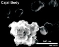 Cajal body EM.jpg 600 × 487; 29 KB
 Cardiac muscle EM01.jpg 1,072 × 735; 231 KB
 Cardiac muscle EM02.jpg 1,072 × 735; 224 KB
 Cardiac muscle EM03.jpg 849 × 615; 135 KB
 Cardiac muscle EM04.jpg 1,000 × 680; 191 KB
 Cardiac Muscle EM05.jpg 992 × 733; 158 KB
 Cartilage em01.jpg 800 × 551; 176 KB
 Collagen EM01.jpg 3,839 × 2,979; 7.77 MB
 Coxsackie B4 virus.jpg 400 × 266; 14 KB
 Cytomegalovirus infected spermatozoa EM01.jpg 990 × 991; 204 KB
 Cytomegalovirus infected spermatozoa.jpg 1,000 × 1,260; 324 KB
 Cytomegalovirus virions EM.jpg 911 × 987; 212 KB
 Cytoplasmic lattices in GV oocyte cytoplasm.jpg 1,048 × 846; 291 KB
 Cytoplasmic lattices in oocytes and two-cell embryos.jpg 753 × 1,000; 226 KB
 Desmosome 02.jpg 600 × 450; 74 KB
 Endotheliochorial placenta EM01.jpg 600 × 602; 134 KB
 Epithelial junctions EM01.jpg 658 × 1,000; 232 KB
 Epithelial junctions EM02.jpg 900 × 1,000; 288 KB
 Epithelial junctions EM03.jpg 1,291 × 1,000; 344 KB
 Erythrocyte and lymphocyte SEM01.jpg 800 × 522; 74 KB
 Erythrocyte and lymphocyte SEM02.jpg 800 × 522; 78 KB
 Erythrocyte and lymphocyte SEM03.jpg 800 × 522; 80 KB
 Fawcett1975 fig31.jpg 1,280 × 403; 128 KB
 Fawcett1975 fig34.jpg 1,280 × 1,746; 506 KB
 Haemomonochorial human placenta EM01.jpg 792 × 775; 79 KB
 Haemomonochorial placenta EM01.jpg 600 × 602; 111 KB
 Hepatitis A virus.jpg 700 × 537; 90 KB
 Hepatitis B virus.jpg 700 × 1,030; 123 KB
 Hepatitis E virus.jpg 700 × 464; 61 KB
 Herpes virus.jpg 320 × 240; 22 KB
 HertigAdams1967 fig01-4.jpg 1,691 × 2,353; 390 KB
 HertigAdams1967 fig01.jpg 531 × 547; 33 KB
 HertigAdams1967 fig02.jpg 531 × 547; 32 KB
 HertigAdams1967 fig03.jpg 518 × 545; 29 KB
 HertigAdams1967 fig04.jpg 1,628 × 1,714; 383 KB
 HertigAdams1967 fig25.jpg 1,637 × 1,452; 256 KB
 Human corpus luteum - light-and-electron-micrograph.jpg 936 × 711; 208 KB
 Human embryo skin 24 week EGA.jpg 596 × 939; 165 KB
 Human embryo skin 8-9 week EGA desmosomes.jpg 800 × 198; 40 KB
 Human embryo skin 8-9 week EGA.jpg 657 × 872; 188 KB
 Human embryo skin 9-11 week EGA.jpg 623 × 804; 176 KB
 Human immunodeficiency virus.jpg 320 × 240; 25 KB
 Human lung inter-alveolar septum em01.jpg 785 × 589; 168 KB
 Human oocyte em01.jpg 600 × 589; 65 KB
 Human ovary follicle basement membrane EM01.jpg 558 × 697; 100 KB
 Human ovary follicles light and electron microscopy 01.jpg 586 × 1,080; 225 KB
 Human papilloma virus.jpg 961 × 721; 218 KB
 Human pronuclear stage EM02.jpg 639 × 1,000; 194 KB
 Human pronuclear stage EM022.jpg 1,100 × 705; 225 KB
 Human pronuclear stage EM03-05.jpg 982 × 439; 104 KB
 Human pronuclear stage EM06.jpg 907 × 1,000; 245 KB
 Human pronuclear stage EM07.jpg 357 × 509; 52 KB
 Human pronuclear stage EM08.jpg 359 × 513; 56 KB
 Human pronuclear stage EM09.jpg 361 × 506; 53 KB
 Human pronuclear stage EM10.jpg 630 × 781; 140 KB
 Human pronuclear stage EM11.jpg 625 × 768; 130 KB
 Human pronuclear stage EM12.jpg 998 × 777; 265 KB
 Human pronuclear stage EM13.jpg 998 × 771; 247 KB
 Human pronuclear stage EM14-16.jpg 1,013 × 459; 141 KB
 Human pronuclear stage EM17.jpg 1,104 × 504; 156 KB
 Human pronuclear stage EM18.jpg 477 × 541; 66 KB
 Human pronuclear stage EM19.jpg 478 × 534; 67 KB
 Human pronuclear stage EM20.jpg 1,114 × 762; 237 KB
 Human pronuclear stage EM21.jpg 366 × 587; 58 KB
 Human pronuclear stage EM22.jpg 366 × 581; 59 KB
 Human pronuclear stage EM25.jpg 1,013 × 782; 201 KB
 Human pronuclear stage EM26.jpg 1,010 × 784; 187 KB
 Human pronuclear stage EM27.jpg 997 × 777; 227 KB
 Human pronuclear stage EM28.jpg 993 × 774; 239 KB
 Human pronuclear stage EM29.jpg 967 × 763; 183 KB
 Human pronuclear stage EM30.jpg 955 × 853; 192 KB
 Human sperm pathologies EM01.jpg 761 × 759; 148 KB
 Human sperm pathology EM02.jpg 800 × 256; 22 KB
 Human spermatid electron micrograph.jpg 619 × 918; 206 KB
 Human spermatid EM01.jpg 1,000 × 762; 162 KB
 Human spermatid EM02.jpg 1,000 × 762; 186 KB
 Human spermatozoa nucleus EM01.jpg 600 × 476; 27 KB
 Human spermatozoa nucleus EM02.jpg 597 × 476; 52 KB
 Human spermatozoa nucleus EM03.jpg 600 × 475; 44 KB
 Human-spermatozoa EM01.jpg 1,000 × 204; 26 KB
 Lassa virus.jpg 320 × 278; 28 KB
 Leydig cell PMID13693345 EM02.jpg 1,359 × 957; 341 KB
 Leydig cell PMID13693345 EM03.jpg 1,359 × 957; 325 KB
 Listeria-bacterium.jpg 320 × 240; 15 KB
 Liver histology EM01.jpg 1,028 × 708; 141 KB
 Liver histology EM02.jpg 1,028 × 707; 154 KB
 Lutein cell glycogen granule em01.jpg 1,149 × 749; 169 KB
 Lutein cell lipid and glycogen em01.jpg 1,156 × 828; 149 KB
 Lutein cell lipid and glycogen em02.jpg 1,109 × 796; 227 KB
 Lymphocyte rosettes EM01-06.jpg 1,364 × 2,100; 334 KB
 Lymphocyte rosettes EM01.jpg 661 × 665; 58 KB
 Lymphocyte rosettes EM012.jpg 618 × 661; 59 KB
 Lymphocyte rosettes EM02.jpg 661 × 665; 62 KB
 Lymphocyte rosettes EM03.jpg 661 × 665; 51 KB
 Lymphocyte rosettes EM04.jpg 661 × 665; 53 KB
 Lymphocyte rosettes EM05.jpg 661 × 665; 55 KB
 Malaria and red blood cell em.jpg 500 × 536; 82 KB
 Marburg virus.jpg 320 × 240; 15 KB
 Measles virus.jpg 700 × 535; 91 KB
 Merkel cell EM 01.jpg 984 × 738; 209 KB
 Merkel cell EM 02.jpg 984 × 685; 166 KB
 MERS-CoV EM1.jpg 537 × 537; 68 KB
 Mitochondria EM01.jpg 640 × 480; 96 KB
 Model capacitation-induced acrosome docking to sperm membrane.jpg 600 × 489; 73 KB
 Monocyte EM01.jpg 923 × 1,000; 221 KB
 Mouse antral follicle 01.jpg 932 × 1,095; 374 KB
 Mouse antral follicle.jpg 600 × 705; 168 KB
 Mouse follicle in vitro 02.jpg 600 × 701; 146 KB
 Mouse follicle in vitro.jpg 600 × 701; 170 KB
 Mouse heart primary cilia 01.jpg 1,200 × 908; 415 KB
 Mouse neonatal ovary oocyte EM01.jpg 677 × 1,000; 266 KB
 Mouse neonatal ovary oocyte EM02.jpg 790 × 792; 179 KB
 Mouse neonatal ovary oocyte EM03.jpg 790 × 792; 187 KB
 Mouse neonatal ovary oocyte EM04.jpg 790 × 792; 235 KB
 Mouse neonatal ovary oocyte EM05.jpg 790 × 792; 217 KB
 Mouse neonatal ovary oocyte EM06.jpg 790 × 792; 163 KB
 Mouse neonatal ovary oocyte EM07.jpg 790 × 792; 183 KB
 Mouse oocyte and zona pellucida EM01.jpg 1,200 × 1,200; 485 KB
 Mouse oocyte and zona pellucida EM01a.jpg 800 × 800; 234 KB
 Mouse oocyte and zona pellucida EM01b.jpg 600 × 600; 133 KB
 Mouse oocyte and zona pellucida EM01c.jpg 400 × 400; 60 KB
 Mouse oocyte balbini body EM01.jpg 695 × 700; 155 KB
 Mouse placenta blood vessel EM01.jpg 1,000 × 739; 132 KB
 Mouse primitive node cilia.jpg 592 × 981; 130 KB
 Mouse renal podocyte EM01.jpg 1,000 × 1,338; 366 KB
 Mouse renal podocyte EM02.jpg 1,000 × 666; 155 KB
 Mouse- cerebellum axons.jpg 600 × 695; 129 KB
 Mouse- spinal cord axons.jpg 600 × 693; 127 KB
 Mouse- zona pellucida 01.jpg 800 × 430; 83 KB
 Mouse-oocyte-d.jpg 790 × 792; 217 KB
 Mouse-optic nerve axons.jpg 600 × 693; 126 KB
 Mouse-sciatic nerve Schwann cell.jpg 957 × 1,050; 339 KB
 Mumps virus.jpg 700 × 938; 221 KB
 Muscle satellite cell EM01.jpg 1,000 × 804; 142 KB
 Muscle satellite cell EM02.jpg 1,000 × 804; 153 KB
 Mycobacterium-tuberculosis.jpg 320 × 240; 18 KB
 Mycoplasma-pneumoniae.jpg 320 × 240; 31 KB
 Neonatal human pulmonary neuroendocrine cell EM01.jpg 836 × 1,200; 405 KB
 Nephron EM01.jpg 1,909 × 1,280; 219 KB
 Nephron EM02.jpg 1,271 × 1,280; 210 KB
 Nephron EM11.jpg 2,983 × 2,000; 398 KB
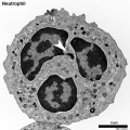 Neutrophil EM01.jpg 999 × 1,000; 402 KB
 Pancreas acinar cell em01.jpg 1,280 × 928; 496 KB
 Parvovirus 01.jpg 1,200 × 796; 275 KB
 Pig sperm capacitation 01.jpg 1,000 × 840; 204 KB
 Pig sperm capacitation 02.jpg 600 × 504; 82 KB
 Pituitary histology 010.jpg 1,005 × 961; 249 KB
 Placental imaging 03A.jpg 800 × 738; 181 KB
 Placental trophospongium.jpg 567 × 344; 94 KB
 Plasma cell EM06.jpg 595 × 600; 84 KB
 Respiratory Epithelium EM01a.jpg 800 × 584; 157 KB
 Respiratory Epithelium EM01b.jpg 220 × 161; 16 KB
 Respiratory Epithelium EM02.jpg 1,200 × 1,005; 483 KB
 Respiratory Epithelium EM02a.jpg 800 × 670; 237 KB
 Respiratory Epithelium EM02b.jpg 220 × 184; 19 KB
 Rotavirus.jpg 700 × 540; 64 KB
 Rubella virus 01.jpg 1,200 × 896; 352 KB
 Rubella virus 02.jpg 700 × 923; 135 KB
 Rubella virus 03.jpg 1,200 × 802; 224 KB
 Rubella virus.jpg 210 × 158; 5 KB
 Spermatozoa tail EM01.jpg 932 × 613; 75 KB
 Staphylococcus-aureus.jpg 320 × 240; 28 KB
 T and B lymphocytes EM09.jpg 1,196 × 627; 137 KB
 T and B lymphocytes EM10.jpg 1,196 × 677; 155 KB
 T2 lymphocyte EM13.jpg 781 × 795; 177 KB
 T2 lymphocyte EM14.jpg 728 × 771; 156 KB
 Treponema-pallidum.jpg 320 × 240; 21 KB
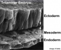 Trilaminar embryo.jpg 432 × 359; 32 KB
 Varicella zoster virus.jpg 800 × 600; 102 KB
 Vascular Sinus EM01.jpg 1,200 × 878; 279 KB
 Vascular Sinus EM01a.jpg 800 × 585; 141 KB
 Vascular Sinus EM01b.jpg 220 × 161; 15 KB
 Venule microvessel EM.jpg 600 × 626; 91 KB
 West Nile virus EM01.jpg 700 × 538; 114 KB
 Zamboni1966 fig02.jpg 1,556 × 1,000; 397 KB
 Zamboni1966 fig06.jpg 1,280 × 1,158; 419 KB
 Zamboni1966 fig20.jpg 1,526 × 1,000; 429 KB
 Zamboni1966 fig29.jpg 1,280 × 1,003; 302 KB
 Zika virus TEM01.jpg 800 × 800; 212 KB


