File:Sea urchin SEM01.jpg
From Embryology
Size of this preview: 800 × 570 pixels. Other resolution: 1,000 × 712 pixels.
Original file (1,000 × 712 pixels, file size: 95 KB, MIME type: image/jpeg)
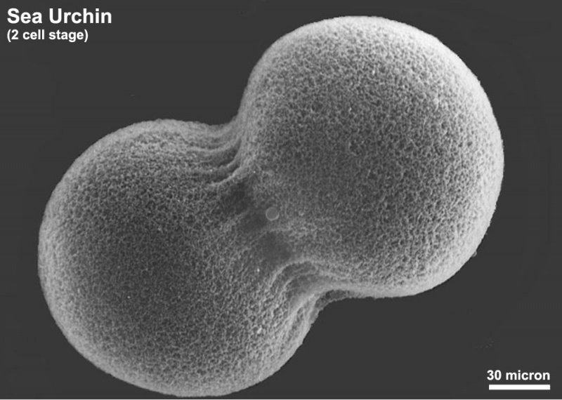
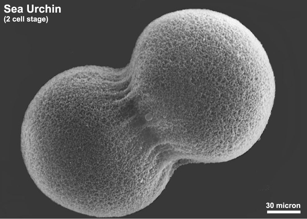
Sea Urchin (2 cell stage)
Scanning electron microscope image of Lytechinus pictus [sea urchin] embryo at the 2-cell stage. Fertilization envelope has been removed to reveal the cells covered with a dense meshwork of the hyaline layer, in which microvilli are embedded.
Class Echinoidea - Superorder Echinacea - OrderTemnopleuroida - Lytechinus pictus
- Links: Sea Urchin Development
JEOL 35C SEM Evelyn Spiegel, Louisa Howard
File history
Yi efo/eka'e gwa ebo wo le nyangagi wuncin ye kamina wunga tinya nan
| Gwalagizhi | Nyangagi | Dimensions | User | Comment | |
|---|---|---|---|---|---|
| current | 14:22, 1 Haziran 2011 | | 1,000 × 712 (95 KB) | S8600021 (talk | contribs) | ==Sea Urchin (2 cell stage)== Scanning electron microscope image of Lytechinus pictus [sea urchin] embryo at the 2-cell stage. Fertilization envelope has been removed to reveal the cells covered with a dense meshwork of the hyaline layer, in which microv |
You cannot overwrite this file.
File usage
The following 2 pages use this file: