File:Liver- Kupffer cell and reticular fibre.jpg
From Embryology

Size of this preview: 450 × 600 pixels. Other resolution: 600 × 800 pixels.
Original file (600 × 800 pixels, file size: 49 KB, MIME type: image/jpeg)
Liver Histology
Liver, rabbit - trichrome & carbon and Liver - reticulin The first slide will allow you to identify the macrophages which adhere to the wall of the liver sinusoids. They are represented by the accumulations of small brown/black dots, the carbon particles ingested by the macrophages.
Source: Blue Histology - Accessory Digestive Glands Liv41re.jpg
http://www.lab.anhb.uwa.edu.au/mb140/CorePages/Liver/liver.htm#LIVER
File history
Yi efo/eka'e gwa ebo wo le nyangagi wuncin ye kamina wunga tinya nan
| Gwalagizhi | Nyangagi | Dimensions | User | Comment | |
|---|---|---|---|---|---|
| current | 13:04, 9 March 2018 | | 600 × 800 (49 KB) | Z8600021 (talk | contribs) | updated size and contrast |
| 09:39, 22 December 2010 | 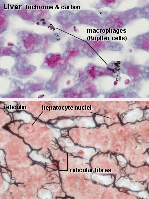 | 300 × 400 (45 KB) | S8600021 (talk | contribs) | ==Liver Histology== Liver, rabbit - trichrome & carbon and Liver - reticulin The first slide will allow you to identify the macrophages which adhere to the wall of the liver sinusoids. They are represented by the accumulations of small brown/black dots, |
You cannot overwrite this file.