File:Fetal 10wk urogenital 3.jpg
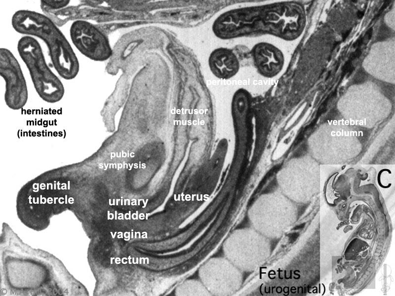
Fetal_10wk_urogenital_3.jpg (800 × 600 pixels, file size: 107 KB, MIME type: image/jpeg)
Human Fetus
female, 10 week, 40 mm CRL, early fetal, sagittal section, pelvic region
This stage of development is after the embryonic period (up to week 8) but still only 2 weeks into early fetal development.
Section A is the most sagittal (lateral towards right) of all sections, plane B, C and D move towards the midline.
Original file name: H10wkUrogenCL.jpg
Image Source: UNSW Embryology http://embryology.med.unsw.edu.au/wwwhuman/Hum10wk/HumUrogen.htm
Related Images: Pelvic region sections progressively lateral towards midline - most lateral | lateral | medial | midline
File history
Yi efo/eka'e gwa ebo wo le nyangagi wuncin ye kamina wunga tinya nan
| Gwalagizhi | Nyangagi | Dimensions | User | Comment | |
|---|---|---|---|---|---|
| current | 19:04, 28 Mayıs 2011 | | 800 × 600 (107 KB) | S8600021 (talk | contribs) | |
| 23:57, 20 September 2009 | 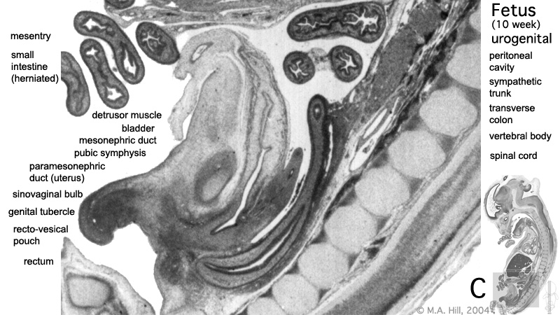 | 800 × 450 (135 KB) | S8600021 (talk | contribs) | These are images from an early fetus (female, 10 week, 40 mm). This stage of development is after the embryonic period (up to week 8) but still only 2 weeks into early fetal development. Section A is the most sagittal (lateral towards right) of all sectio |
You cannot overwrite this file.
File usage
The following 19 pages use this file:
- 2009 Lecture 15
- 2010 Lab 8
- 2010 Lecture 15
- 2011 Lab 12 - Embryo to Fetus
- 2011 Lab 5 - Fetal
- 2011 Lab 8 - Fetal
- ANAT2241 Urinary System
- ANAT2341 Lab 11 - Embryo to Fetus
- ANAT2341 Lab 12 - Embryo to Fetus
- ANAT2341 Lab 5 - Fetal
- ANAT2341 Lab 8 - Fetal
- BGDB Sexual Differentiation - Fetal
- Fetal Development - 10 Weeks
- Genital - Female Development
- Lecture - Renal Development
- Renal System - Fetal
- Renal System Development
- Renal System Histology
- Urinary Bladder Development


