File:Spleen histology 03.jpg
Spleen_histology_03.jpg (450 × 600 pixels, file size: 83 KB, MIME type: image/jpeg)
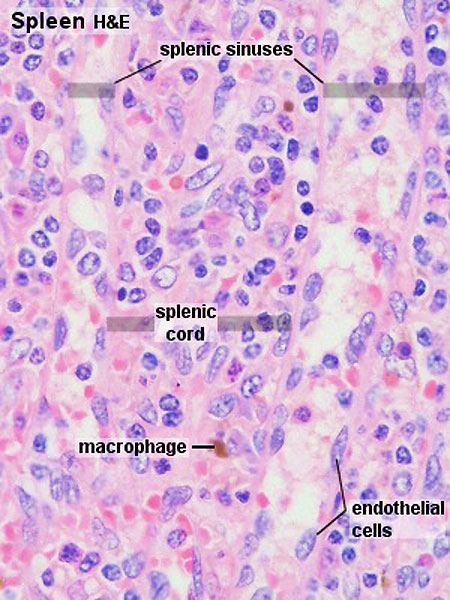
Spleen Histology - Cords and Sinuses
Blood cells which are emptied into the splenic cords re-enter the blood vessels through the endothelium of the sinusoids. The endothelial cells are elongated (in cross section they may appear cuboidal) and oriented along the long axis of the sinusoids. The endothelium of the sinusoids has no junctional complexes and its basement membrane is incomplete (forming narrow circular bands around the endothelial cells with large intervening fenestrations). Macrophages ingest aged erythrocytes, platelets and other particulate matter as they pass through the splenic cords.
The sinusoids continue into the veins of the pulp, which empty into thin-walled trabecular veins, which eventually coalesce to form the splenic vein.
Links: Histology | Histology Stains | Blue Histology images copyright Lutz Slomianka 1998-2009. The literary and artistic works on the original Blue Histology website may be reproduced, adapted, published and distributed for non-commercial purposes. See also the page Histology Stains.
Cite this page: Hill, M.A. (2026, October 7) Embryology Spleen histology 03.jpg. Retrieved from https://embryology.med.unsw.edu.au/embryology/index.php/File:Spleen_histology_03.jpg
- © Dr Mark Hill 2026, UNSW Embryology ISBN: 978 0 7334 2609 4 - UNSW CRICOS Provider Code No. 00098G
Original File name: Spl42he.jpg
File history
Yi efo/eka'e gwa ebo wo le nyangagi wuncin ye kamina wunga tinya nan
| Gwalagizhi | Nyangagi | Dimensions | User | Comment | |
|---|---|---|---|---|---|
| current | 20:30, 22 February 2012 | | 450 × 600 (83 KB) | Z8600021 (talk | contribs) | |
| 15:55, 21 February 2011 |  | 300 × 400 (51 KB) | S8600021 (talk | contribs) | ==Spleen Histology== Original File name: Spl42he.jpg {{Blue Histology}} Category:Spleen Category:Endocrine Category:Histology Category:Immune |
You cannot overwrite this file.


