File:Mouse Wnt signaling 01.jpg
Mouse_Wnt_signaling_01.jpg (600 × 450 pixels, file size: 156 KB, MIME type: image/jpeg)
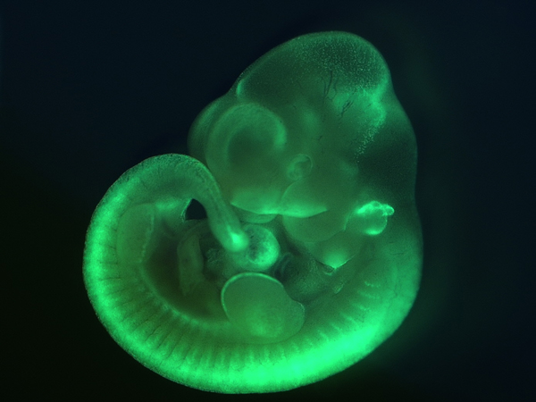
Mouse Wnt/beta-Catenin Signaling
The development of a novel mouse transgenic line allows the study of Wnt signaling activity in living embryos at single-cell resolution. Transgenic mice express a reporter comprising GFP fused to human histone H2B under the control of Wnt/ß-catenin signaling pathway response elements. Expression of GFP mimics canonical Wnt/ß -catenin signaling activity. At embryonic day 10.5, as shown by live imaging, GFP expression is observed in the neural tube, somites, heart, nasal and pharyngeal region (GFP; green)
Original File Name: 201012large.jpg http://www.biomedcentral.com/graphics/journal/imageofthemonth/1005/201012large.jpg
Reference
<pubmed>21176145</pubmed>| BMC Dev Biol.
© 2010 Ferrer-Vaquer et al. ; licensee BioMed Central Ltd. This is an open access article distributed under the terms of the Creative Commons Attribution License (http://creativecommons.org/licenses/by/2.0), which permits unrestricted use, distribution, and reproduction in any medium, provided the original work is properly cited.
File history
Yi efo/eka'e gwa ebo wo le nyangagi wuncin ye kamina wunga tinya nan
| Gwalagizhi | Nyangagi | Dimensions | User | Comment | |
|---|---|---|---|---|---|
| current | 15:07, 28 December 2010 | | 600 × 450 (156 KB) | S8600021 (talk | contribs) | ==Mouse Wnt/beta-Catenin Signaling== The development of a novel mouse transgenic line allows the study of Wnt signaling activity in living embryos at single-cell resolution. Transgenic mice express a reporter comprising GFP fused to human histone H2B und |
You cannot overwrite this file.
File usage
The following page uses this file:


