File:Licata1954 fig03.jpg

Original file (1,000 × 708 pixels, file size: 92 KB, MIME type: image/jpeg)
=Fig. 3 Dorsal aspect of heart of 31.5 mm embryo
Showing the pericardial reflections where the heart was cut from the dorsal mediastinal wall. The fold of Marshall has been cut to show the transverse pericardial sinus (arrow). The course of the oblique vein within the fold of Marshall is indicated by interrupted lines.
(Reconstruction X 100, illustration X 34.)
Reference
Licata RH. The human embryonic heart in the ninth week. (1954) Amer. J Anat., 94: 73-125. PMID 13124266
Cite this page: Hill, M.A. (2026, October 4) Embryology Licata1954 fig03.jpg. Retrieved from https://embryology.med.unsw.edu.au/embryology/index.php/File:Licata1954_fig03.jpg
- © Dr Mark Hill 2026, UNSW Embryology ISBN: 978 0 7334 2609 4 - UNSW CRICOS Provider Code No. 00098G
File history
Yi efo/eka'e gwa ebo wo le nyangagi wuncin ye kamina wunga tinya nan
| Gwalagizhi | Nyangagi | Dimensions | User | Comment | |
|---|---|---|---|---|---|
| current | 11:42, 5 March 2017 | | 1,000 × 708 (92 KB) | Z8600021 (talk | contribs) | |
| 11:41, 5 March 2017 | 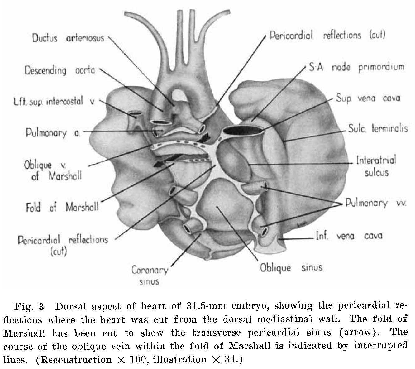 | 1,332 × 1,184 (234 KB) | Z8600021 (talk | contribs) |
You cannot overwrite this file.
File usage
The following 4 pages use this file:


