File:Frazer1914 fig01.jpg
From Embryology
Size of this preview: 800 × 468 pixels. Other resolution: 1,000 × 585 pixels.
Original file (1,000 × 585 pixels, file size: 113 KB, MIME type: image/jpeg)
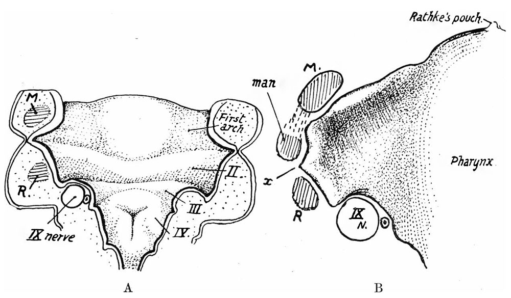
Fig. 1 gives in outline the shape of the pharyngeal cavity in a 12-mm. embryo, and illustrates this description, showing how each broad and open recess is floored by the outer parts of the first two arches, how their grooves lie behind them and end externally in deeper lateral pouches, and how the third arch, lying between the second and third pouches, necessarily forms the short posterior boundary of the recess.
File history
Yi efo/eka'e gwa ebo wo le nyangagi wuncin ye kamina wunga tinya nan
| Gwalagizhi | Nyangagi | Dimensions | User | Comment | |
|---|---|---|---|---|---|
| current | 05:58, 9 Ocak 2017 | | 1,000 × 585 (113 KB) | Z8600021 (talk | contribs) | |
| 05:53, 9 Ocak 2017 |  | 1,442 × 903 (208 KB) | Z8600021 (talk | contribs) |
You cannot overwrite this file.
File usage
The following 3 pages use this file:


