Talk:Draft 2016
From Embryology
Lymph Nodes
Lymph Node Anatomy
| Lymph Node Anatomy | |
|---|---|
Mesenteric lymph nodes |

|

Lymph Node Functions
| Lymph Node Functions | |
|---|---|
|

|
Lymph Node Structure
| Lymph Node Structure | |||||||
|---|---|---|---|---|---|---|---|

Simplified structure |

Lymphocyte (T and B) Traffic
| ||||||

|
Lymph pathway
Watch T and B Lymphocytes Move
| ||||||

| |||||||
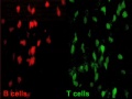
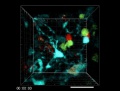
Lymph Node Histology
| Lymph Node Histology | ||||||||||||
|---|---|---|---|---|---|---|---|---|---|---|---|---|

Connective Tissue
Links: Immunobiology - Figure 1.8. Organization of a lymph node |


Diffuse Lymphatic Tissue

Alimentary canal, respiratory passage and urogenital tract.
- BALT - Bronchus Associated Lymphoid Tissue or GALT - Gut Associated Lymphatic Tissue
- Not enclosed by a connective tissue capsule
- Located in subepithelial tissue - lamina propria
- Diffuse lymphatic tissue + nodules
- Reactive - enlarge when activated (by antigen)
Lymphocytes
- travel to nodes and back again
- proliferation and differentiation
Lymph Nodules
- Organized concentrations of lymphocytes
- No capsule, covered by epithelia
- Nodules are also the unit structure seen in a node
- Oval concentrations in meshwork of reticular cells
Nodule States
- Primary Nodule - Mainly small lymphocytes
- Secondary Nodule
- Central pale region (germinal centre) - Effector cells and macrophages
- Dark outer ring (small lymphocytes)
Gastrointestinal Tract
- Oropharynx - Tonsils
- Distal small intestine (ilieum) - Peyer’s Patches
- Appendix, cecum
Mucosal Associated Lymphoid Tissues

|
Anatomical location - Palatine (tonsils), Lingual and Pharyngeal ( adenoids ) Ring of oral adenoid tissue:
|
Palatine Tonsils


- the "tonsils", lateral wall of oropharynx
- covered by stratified squamous epithelium
- numerous crypts (10-20) infolds of surface epithelium
- Afferent lymph vessels absent
- Efferent lymph vessels are present
Lingual Tonsils
- lamina propria root of tongue
- covered by stratified squamous epithelium
- salivary glands and skeletal muscle are directly adjacent
Pharyngeal Tonsils
- adenoids or nasopharyngeal tonsils, upper posterior part of throat
- covered by a pseudostratified ciliated epithelium with goblet cells
Peyer's Patch
- located in the ileum

|

|

|
| Peyers patches (ileocolonoscopy) | Peyer's Patch (histology) | microfold cells or M-cells (transport gut lumen organisms and particles to immune cells across the epithelial barrier). |
| About Peyer's Patch |
|---|
External Links Notice - The dynamic nature of the internet may mean that some of these listed links may no longer function. If the link no longer works search the web with the link text or name. Links to any external commercial sites are provided for information purposes only and should never be considered an endorsement. UNSW Embryology is provided as an educational resource with no clinical information or commercial affiliation.
|

