File:Uterus proliferative phase.jpg
Uterus_proliferative_phase.jpg (400 × 533 pixels, file size: 52 KB, MIME type: image/jpeg)
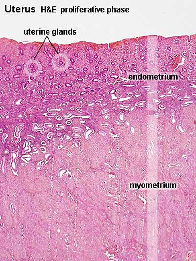
Uterus Histology - Proliferative Phase
Histology showing endometrium and myometrium during proliferative phase of menstrual cycle H&E stain
Image Source: UWA Blue Histology http://www.lab.anhb.uwa.edu.au/mb140/CorePages/FemaleRepro/femalerepro.htm#Uterus
Links: Histology | Histology Stains | Blue Histology images copyright Lutz Slomianka 1998-2009. The literary and artistic works on the original Blue Histology website may be reproduced, adapted, published and distributed for non-commercial purposes. See also the page Histology Stains.
Cite this page: Hill, M.A. (2026, October 3) Embryology Uterus proliferative phase.jpg. Retrieved from https://embryology.med.unsw.edu.au/embryology/index.php/File:Uterus_proliferative_phase.jpg
- © Dr Mark Hill 2026, UNSW Embryology ISBN: 978 0 7334 2609 4 - UNSW CRICOS Provider Code No. 00098G
File history
Yi efo/eka'e gwa ebo wo le nyangagi wuncin ye kamina wunga tinya nan
| Gwalagizhi | Nyangagi | Dimensions | User | Comment | |
|---|---|---|---|---|---|
| current | 13:57, 2 February 2012 | | 400 × 533 (52 KB) | S8600021 (talk | contribs) | increase image size |
| 11:21, 3 August 2009 |  | 300 × 400 (66 KB) | MarkHill (talk | contribs) | Uterus proliferative phase Histology showing endometrium and myometrium during proliferative phase of menstrual cycle H&E stain Image Source: UWA Blue Histology http://www.lab.anhb.uwa.edu.au/mb140/CorePages/FemaleRepro/femalerepro.htm#Uterus |
You cannot overwrite this file.
File usage
The following 10 pages use this file:


