File:Peyer's patch 02.jpg
From Embryology
No higher resolution available.
Peyer's_patch_02.jpg (450 × 600 pixels, file size: 69 KB, MIME type: image/jpeg)
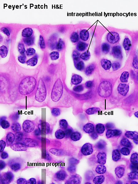
Peyer's Patch
|
|
- Immune Images: Oesophagus MALT | Colon MALT | Peyer's patch overview | Peyer's patch detail | Cartoon - IEL development | Cartoon - IEL function | Cartoon - IEL differentiation | Mesenteric Lymph Nodes overview | Palatine Tonsil | Tonsil | Immune System Development
- ↑ <pubmed>21681197</pubmed>
Links: Histology | Histology Stains | Blue Histology images copyright Lutz Slomianka 1998-2009. The literary and artistic works on the original Blue Histology website may be reproduced, adapted, published and distributed for non-commercial purposes. See also the page Histology Stains.
Cite this page: Hill, M.A. (2024, June 14) Embryology Peyer's patch 02.jpg. Retrieved from https://embryology.med.unsw.edu.au/embryology/index.php/File:Peyer%27s_patch_02.jpg
- © Dr Mark Hill 2024, UNSW Embryology ISBN: 978 0 7334 2609 4 - UNSW CRICOS Provider Code No. 00098G
File history
Click on a date/time to view the file as it appeared at that time.
| Date/Time | Thumbnail | Dimensions | User | Comment | |
|---|---|---|---|---|---|
| current | 10:05, 24 February 2012 | | 450 × 600 (69 KB) | Z8600021 (talk | contribs) | |
| 08:23, 24 December 2010 |  | 300 × 400 (41 KB) | S8600021 (talk | contribs) | Peyer's patch 01.jpg Pey102he.jpg |
You cannot overwrite this file.
File usage
The following 3 pages use this file: